
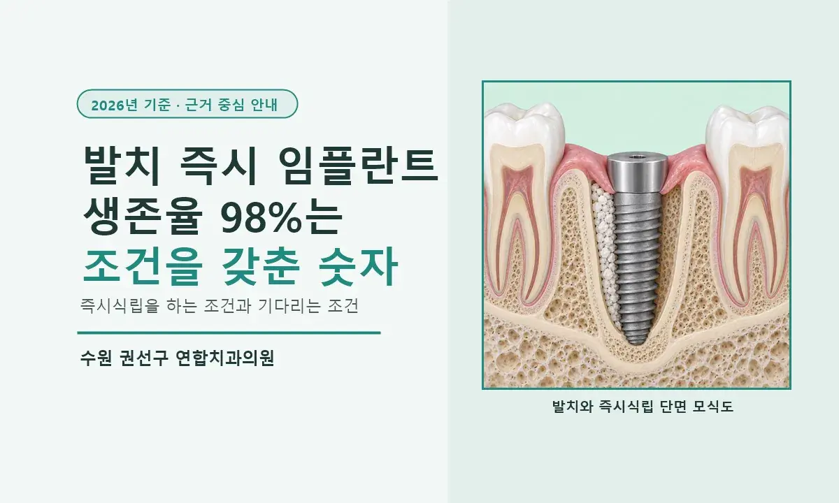
An immediate implant after extraction (immediate placement) places the implant in the same site on the day the tooth is removed. It removes the 2 to 3 months of waiting after extraction but does not shorten the 3 to 6 months it takes to bond to the bone. The 2-year estimated survival of 98.4% is a value calculated by pooling many studies, and in direct comparison studies, immediate placement had more early failures. We have laid out in numbers the conditions under which Yeonhap Dental Clinic places immediately and the conditions under which we wait.
Who can have an immediate implant after extraction in Suwon?
The idea that placing the implant on the day of extraction means you do not have to come to the clinic twice is only half true.
[Key Answer]
An immediate implant after extraction (immediate placement) places the implant in the extraction site on the same day, removing the 2 to 3 months of waiting after extraction but not the 3 to 6 months of bonding to the bone. A systematic review pooling 46 prospective studies reports a 2-year estimated survival of 98.4% (Lang 2012), but in studies directly comparing immediate and delayed placement, immediate placement had more early failures (Cosyn 2019). Yeonhap Dental Clinic decides whether to place on the same day by looking at four things: removal of infection, fixation beneath the extraction socket, distance to the maxillary sinus and nerve canal, and the gum in the front teeth.
[Summary in 4 Lines]
· After a tooth is removed, the width of the jawbone shrinks by an average of 3.79 mm (29 to 63%) within 6 months, and the cheek-side wall disappears first (Tan 2012). Immediate placement removes this wait but does not prevent the resorption itself.
· The 2-year estimated survival of 98.4% for immediate placement (Lang 2012) and the 94.9% immediate versus 98.9% delayed in a direct comparison (Cosyn 2019) cannot be compared directly because the study designs and follow-up periods differ. In a subanalysis of studies that used antibiotics after surgery, no significant difference in survival between the two methods was observed, but antibiotics alone cannot explain it.
· Yeonhap Dental Clinic divides cases into same-day placement and placement after waiting, and aims for insertion torque of 30 to 35 Ncm, obtaining fixation in the original bone 2 to 3 mm below the floor of the socket. If fixation is barely achieved at 10 Ncm or less, we do not place that day and wait at least 2 months for front teeth and 3 months for molars before placing.
· After a flapless extraction and curettage, if torque is 30 Ncm or more we cover with a healing abutment, and if lower with a low healing abutment or cover screw; we place 5 to 7 mm below the gum surface, and add bone graft material if the gap between the implant and bone exceeds 2 mm.
Written by Dr. Ahn Seong-jin, Chief Director, Yeonhap Dental Clinic
[Table of Contents]
· How much bone is lost after a tooth is removed, and where does it start?
· How should the 98% survival rate for immediate placement be read?
· How do same-day placement and placement after waiting differ?
· Under what conditions does Yeonhap Dental Clinic place immediately, and when does it wait?
· Where and how is primary stability obtained in an extraction socket?
· How are immediate implants placed after extraction of front teeth and molars?
· Why is a 2 mm gap between the implant and bone the criterion?
· How do I get by with temporary teeth and the waiting period after immediate placement?
An immediate implant after extraction (immediate implant placement) places the implant on the same day in the extraction socket (the bone hole left where the tooth was). Bone at the extraction site shrinks over time, and even immediate placement does not stop that resorption, so the benefit of immediate placement lies less in preserving bone than in removing the waiting time. The term "immediate placement" can sound like "finished quickly," but what disappears is the time spent waiting for healing after extraction, while the osseointegration period in which the implant bonds to the bone (about 3 months in the lower jaw and 4 to 6 months in the upper jaw) stays the same.
This article lays out, in order, how much bone is lost after extraction, how to read the papers on immediate placement survival, the conditions under which Yeonhap Dental Clinic does and does not place immediately, and the criteria for primary stability, depth, gap, and temporary teeth. This post was written as of September 2026.
How much bone is lost after a tooth is removed, and where does it start?
After a tooth is removed, the width of the jawbone shrinks by an average of 3.79 mm within 6 months, and the cheek-side bone wall disappears first.
In a systematic review pooling 20 studies that followed human extraction sockets, 6 months after extraction the horizontal bone width had shrunk by an average of 3.79 mm and the height by 1.24 mm on the cheek side (Tan 2012). In proportion, the horizontal width shrank by 29 to 63% and the height by 11 to 22%. In a study that reopened implant sites 4 months after extraction and placement, about 56% of the thickness of the cheek-side bone wall and about 30% of the tongue-side wall had resorbed, so the cheek side collapsed much faster (Botticelli 2004).
This resorption does not stop even if an implant is placed right away. In a dog experiment placing immediate implants in extraction sockets, the cheek-side wall shrank similarly to the side without an implant (Araújo 2005). Immediate placement is not surgery that prevents bone resorption; it is surgery that secures the implant position before resorption begins.
That is why Yeonhap Dental Clinic places the implant deeper in immediate placement than in conventional placement. Conventional placement is based on 4 mm below the gum surface, but because the amount of bone that will shrink cannot be calculated in advance in immediate placement, we go down to 5 to 7 mm. This is so that the neck of the implant does not become exposed outside the gum even after the bone has shrunk. The 4 mm standard for conventional placement is covered in What Decides Implant Placement Depth?.
How should the 98% survival rate for immediate placement be read?
The 2-year estimated survival of 98.4% for immediate placement is a value calculated from annual failure rates pooled across studies, and in studies directly comparing immediate and delayed placement, immediate placement had more early failures.
Pooling 46 prospective studies with follow-up of at least 1 year (average follow-up 2.08 years), the annual failure rate of immediate placement is 0.82%, and the converted 2-year estimated survival is 98.4% (Lang 2012). But narrowing to studies that directly compare immediate and delayed placement changes the result. In a meta-analysis pooling 8 studies and 473 implants functioning for 12 to 96 months, survival was 94.9% for immediate placement and 98.9% for delayed placement, and all failures were early failures (Cosyn 2019). However, that meta-analysis rated most of the included studies as having a high risk of bias.
Because the study designs and follow-up periods differ, the figures from the two reviews cannot be compared directly. In the subanalysis of the direct comparison meta-analysis, studies that used antibiotics after surgery showed no significant difference in survival between immediate and delayed placement, but studies that did not use antibiotics also showed only a trend-level difference, so the effect of antibiotics alone cannot explain it. In the 46-study review, of five variables analyzed, only whether antibiotics were prescribed after surgery had a significant effect on survival (Lang 2012).
In a meta-analysis that separately pooled cases placed in infected sockets, if the socket was thoroughly curetted (scraping out inflamed tissue), there was no significant difference in survival from sockets without infection (Lee 2018). However, this is limited evidence made up of 5 comparative studies with no randomized study, and it reflects cases in which curettage was carried through. This is why Yeonhap Dental Clinic waits when pus and swelling are severe and the infection cannot be fully removed.
Comparison of same-day placement and placement after waiting
| Item | Same-day placement | Placement after waiting |
|---|---|---|
| Time of placement | Day of extraction | 2 months after extraction for front teeth, at least 3 months for molars |
| Wait after extraction | None | 2 to 3 months |
| Period of bonding to the bone | About 3 months in the lower jaw, 4 to 6 months in the upper jaw | Same |
| Position of primary stability | Mainly the original bone 2 to 3 mm below the socket floor | Healed bone |
| Bone graft | Simultaneous graft if the gap exceeds 2 mm | The graft amount for the gap may decrease, but a graft may be needed if the cheek-side bone resorbs |
| 2-year estimated survival (Lang 2012) | 98.4% | No comparison value in that study |
| Survival in direct comparison studies (Cosyn 2019, 12 to 96 months) | 94.9% | 98.9% |
| Cheek-side bone | Resorption proceeds similarly even with immediate placement | Bone resorption proceeds during the wait; judged by the remaining bone at the time of placement |
| Yeonhap Dental Clinic's choice | Infection can be curetted, fixation in bone below the socket exceeds 10 Ncm, room from the sinus and nerve canal, front tooth gum maintained | Other cases |
Under what conditions does Yeonhap Dental Clinic place immediately, and when does it wait?
We place immediately when the infection can be fully removed, fixation is achieved in the original bone below the socket floor, there is no maxillary sinus or nerve canal at that depth, and, for a front tooth, the gum has not receded.
The first is infection. After the tooth is removed, we scrape out the inflamed tissue in the socket with curettage, and if we judge that pus and swelling are too severe to remove completely, we do not place that day. This is because the results of studies on infected sockets are also figures for cases in which curettage was thorough.
The second is the bone beneath the socket. Yeonhap Dental Clinic obtains fixation mainly by engaging the screw in the original bone 2 to 3 mm below the socket floor, and if fixation is so poor that insertion torque is 10 Ncm or less and even the cover screw is hard to tighten by hand, we do not place that day. The third is what lies at that depth. If the maxillary sinus (the empty space beside the nose) is close in an upper molar, or the inferior alveolar nerve canal (the large nerve running through the lower jaw) is close in a lower molar, there is a risk of reaching them while securing 2 to 3 mm, so we wait and plan after the bone has healed.
The fourth is the gum in the front teeth. We place most front teeth on the same day unless the gum has already receded from infection. Because the proportion of immediately placed front teeth in which the gum receded by more than 1 mm is reported at a median of 26% (Chen 2014), we wait where the gum has receded. If any one of these four applies, Yeonhap Dental Clinic does not place that day and waits at least 2 months for front teeth and 3 months for molars before placing. The changes that occur in the empty site's bone and neighboring teeth during the wait are summarized in Geumgok-dong Implants.
Where and how is primary stability obtained in an extraction socket?
Primary stability is obtained mainly in the original bone 2 to 3 mm below the socket floor, aiming for an insertion torque of 30 to 35 Ncm.
Primary stability (the force with which the implant grips the bone at the moment of placement) comes mainly from the bone below the floor rather than the widely flared socket walls. Yeonhap Dental Clinic usually drills about 3 mm below the socket floor to engage the tip of the screw in the original bone and aims for an insertion torque of 30 to 35 Ncm. Considering the effect that excessive compression may have on blood flow to the surrounding bone, we do not tighten further to obtain a higher torque. If the torque falls short of 30 Ncm, we leave the implant as placed and cover it with a low healing abutment or cover screw so that no load is applied, and if fixation is barely achieved at 10 Ncm or less, we do not place that day and wait. This torque range is not a figure from the literature but Yeonhap Dental Clinic's clinical standard.
Surgery is done flapless, without cutting the gum. In a meta-analysis of 5 randomized studies comparing flap and flapless surgery in immediate placement, the cheek-side bone was preserved 0.48 mm more with flapless, and there was no difference in survival (Pitman 2023). However, most were studies of single immediate implants in sockets with nearly intact walls followed for 6 to 12 months, so the clinical significance is still limited. It is interpreted that not opening the gum maintains blood flow to the cheek-side bone and reduces resorption.
After placement, if the insertion torque is 30 Ncm or more, we close the entrance of the socket with a healing abutment (a cap that rises above the gum). We use 5.0 mm in diameter for front teeth and premolars and 6.0 mm for molars. Not adding sutures or a barrier membrane over it is Yeonhap Dental Clinic's method, and the study showing that the gap around the implant can fill with new bone (Botticelli 2004) was done with the gum opened and sutured, so it does not directly prove this method. Prescribing 5 days of antibiotics on the day of surgery is also our clinic's policy, referring to a report that whether antibiotics are prescribed after surgery was related to survival (Lang 2012).
How are immediate implants placed after extraction of front teeth and molars?
A front tooth immediate implant is placed on the same day in most cases if the gum has not receded, with the implant set against the palatal side and the cheek-side gap filled with bone graft material.
In front teeth, the cheek-side bone is thin, so this is the site most affected by post-extraction resorption. Yeonhap Dental Clinic places most on the extraction day unless the gum has already receded from infection, but because resorption proceeds even with immediate placement, we prepare through position and gap management. The proportion of immediately placed front teeth in which the gum receded by more than 1 mm was a median of 26% across 8 studies (range 9 to 41%, 1 to 3 years after placement) (Chen 2014), and without keeping to position and gap management, this can result in the front tooth looking longer than its neighbors.
Yeonhap Dental Clinic places the front tooth implant not in the center of the socket but against the palatal wall, angling it so the head sits inside the line connecting the incisal edges of the neighboring teeth. This leaves a gap on the cheek side by necessity, and we fill it with bone graft material to build 2 mm of cheek-side bone thickness. Primary stability, as with molars, is obtained mainly in the original bone 2 to 3 mm below the socket floor, aiming for 30 to 35 Ncm, and for upper front teeth we use a diameter of 3.0 to 4.0 mm and a length of 8.5 mm or more.
We consider a temporary tooth only if insertion torque is 30 Ncm or more, and even then we decide case by case together with other conditions such as the bite and deep bite (front teeth biting deeply). When we do place one, we keep it clearly out of contact with the opposing teeth. If torque falls short of 30 Ncm, we use a flipper (a removable temporary denture) instead of a temporary tooth, and even when we did not place that day, the patient uses a flipper for 2 months after extraction before placement.
A molar immediate implant uses a diameter of 4.0 to 5.0 mm (premolars 4.0 to 4.5 mm, molars 4.5 to 5.0 mm), placed in the bone between the roots or at one root site, wherever fixation can be obtained in the bone 2 to 3 mm below.
A molar socket has two or three roots, so the hole is wide and irregular in shape. In a meta-analysis pooling 20 studies and 1,106 implants, the 1-year survival of immediate molar placement was 96.6% and the success rate 93.3%, with marginal bone loss 0.31 mm greater than in healed bone (Ragucci 2020). In the same analysis, whether the gap was grafted and when chewing was allowed affected the results.
Yeonhap Dental Clinic uses a diameter of 4.0 to 5.0 mm for immediate molar placement. This is because a wide socket needs a thick implant to secure contact with the bone. The bone between the roots (the septum) is preferred, but even if the septum is absent or narrow, we place the implant where fixation can be obtained in the original bone 2 to 3 mm below. For a lower molar, that means the mesiolingual (inner front) root site, and for an upper molar, the palatal (toward the palate) root site. If fixation is 10 Ncm or less, we do not place that day and wait. If it exceeds 10 Ncm but falls short of 30 Ncm, we protect it with a low healing abutment or cover screw. The criteria for molar diameter and length are in Why Front Tooth and Molar Implants Differ in Width, Length, and Number.
Why is a 2 mm gap between the implant and bone the criterion?
Yeonhap Dental Clinic's standard is to fill the gap with bone graft material when it exceeds 2 mm between the implant and the socket wall.
When an implant is placed in an extraction socket, a gap remains between the implant surface and the bone wall. In a study that placed implants with the gum opened, left this gap unfilled, and reopened the site 4 months later, 44 of 52 gaps wider than 3 mm had filled with new bone (Botticelli 2004). A gap can fill with new bone, but in the same study about 56% of the thickness of the cheek-side wall resorbed. The thinner the cheek-side wall, the more easily it collapses inward as it heals.
Yeonhap Dental Clinic adds bone graft material when the gap exceeds 2 mm. In particular, because the front tooth implant is placed against the palatal side, a gap necessarily forms on the cheek side, and we fill it to build 2 mm of cheek-side bone thickness. The graft is done on the same day as placement, and we do not use the approach of placing graft material first and the implant a few months later. The types of bone graft material and fixation methods are covered in Implant Bone Grafting: When and How.
Yeonhap Dental Clinic's criteria for immediate implants after extraction
| Item | Criterion | Reason |
|---|---|---|
| Time of placement | Day of extraction, or after waiting (2 months for front teeth, at least 3 months for molars) | Decided by infection, fixation, maxillary sinus/nerve canal, and the front tooth gum |
| Infection | Immediate only when it can be completely removed by curettage | Survival in infected sockets assumes curettage |
| Position of primary stability | Mainly the original bone 2 to 3 mm below the socket floor | Wide socket walls alone give insufficient fixation |
| Insertion torque | Aim for 30 to 35 Ncm; do not tighten further beyond that | Considering the effect of excessive compression on blood flow to surrounding bone (our clinic's standard) |
| Torque below 30 Ncm | If above 10 Ncm, low healing abutment or cover screw after placement; if 10 Ncm or less, do not place that day | Block loading according to the degree of fixation (our clinic's standard) |
| Maxillary sinus / nerve canal | No immediate placement if securing 2 to 3 mm would reach them | Safety distance comes first |
| Incision | Flapless | 0.48 mm more cheek-side bone preserved reported (Pitman 2023) |
| Socket entrance | If 30 Ncm or more, healing abutment of 5.0 mm (front teeth, premolars) or 6.0 mm (molars) | No sutures or barrier membrane (our clinic's method) |
| Placement depth | 5 to 7 mm from the gum surface | Deeper than the 4 mm of conventional placement, to allow for bone resorption |
| Gap management | Simultaneous bone graft if over 2 mm | Our clinic's standard; small gaps are judged by the state of the cheek-side wall |
| Molar diameter | 4.0 to 5.0 mm, septum or root site | To secure contact surface in a wide socket |
| Temporary tooth | Front teeth on the same day if 30 Ncm or more and the bite allows (out of contact with the opposing tooth), otherwise a flipper. Molars: healing abutment or cover screw | To block loading before bonding to the bone |
| Antibiotics | 5-day prescription on the day of surgery | Our clinic's policy, referring to a report linking antibiotic prescription to survival (Lang 2012) |
| Upper front tooth diameter | 3.0 to 4.0 mm, set against the palatal side | To secure a 2 mm cheek-side gap |
How do I get by with temporary teeth and the waiting period after immediate placement?
For front teeth, if conditions allow, a temporary tooth out of contact with the opposing teeth is placed on the day of placement; for molars, only a healing abutment or cover screw is left and you chew on the other side.
For a front tooth temporary, insertion torque of 30 Ncm or more is the minimum condition, and we decide whether to place it after looking at other conditions such as the bite and deep bite. When we do, we adjust it so it is clearly separated from the opposing teeth, visibly so, so that bite force does not act directly on the temporary tooth. This is because loading before the implant bonds to the bone can lead to failure. If the conditions are not met, we use a flipper (a removable temporary denture) instead of a temporary tooth. For molars, you chew on the other side without a temporary tooth until the bone bonds.
Immediate placement and immediate loading are different terms. Immediate loading means allowing chewing on a temporary tooth from the day of placement, and applies only to cases that meet the conditions when connecting several implants. An immediate temporary tooth for one or two front teeth is a non-chewing temporary prosthesis, and either way the timing of the final prosthesis is the same, after the bone has bonded.
If we did not place that day, for front teeth you manage with a flipper for 2 months after extraction, and for molars you leave the site empty for at least 3 months and chew on the other side. For an upper molar that had sinusitis, we recheck the sinus on CBCT 3 months after extraction before planning. As the socket heals, the amount of graft needed to fill the gap may decrease, but if the cheek-side bone resorbs during the wait, a bone graft may actually be needed. The total treatment period lengthens by this wait, while the bonding period stays the same.
4 things to check when consulting about an immediate implant after extraction
Whether immediate placement is possible is gauged by the four points below and confirmed with the CT and the condition of the socket at your first visit. The actual fixation (insertion torque) is checked during surgery. Information that is helpful to share before your first consultation is summarized in Suwon Immediate Implant After Extraction.
· Whether there is pus, swelling, or a sinus tract around the tooth to be removed, and whether it can be fully removed by curettage
· Whether there is at least 2 to 3 mm of original bone below the socket floor to hold the implant, and whether the maxillary sinus or inferior alveolar nerve canal is reached at that depth
· For a front tooth, whether the gum has already receded and whether cheek-side bone remains
· For a molar, whether CT shows enough bone, in position and amount, between the roots or at one root site to obtain fixation
[Key Takeaways]
✓ Immediate placement removes the 2 to 3 months of waiting after extraction, and does not shorten the 3 to 6 months of bonding to the bone.
✓ The 2-year estimated survival of 98.4% for immediate placement (Lang 2012) and the 94.9% immediate versus 98.9% delayed in a direct comparison study (Cosyn 2019) cannot be compared directly because the designs and follow-up periods differ, and the antibiotic subanalysis alone does not explain the difference.
✓ Yeonhap Dental Clinic aims for 30 to 35 Ncm of fixation in the original bone 2 to 3 mm below the socket floor, covers with a low healing abutment or cover screw if below 30 Ncm, and does not place that day if fixation is 10 Ncm or less.
What I would like to tell you about immediate placement from the treatment room
I think that choosing precisely the days when we should not place immediately matters more than being good at immediate placement. Putting an implant into a socket is not itself difficult. What is difficult is, when fixation is so poor that even the cover screw is hard to tighten by hand, leaving the extraction site empty and telling you to wait at least 2 months for a front tooth or 3 months for a molar and then look again.
I do it anyway. Rather than expecting bone to bond to an implant with almost no fixation, it is ultimately better for the patient to wait, reassess the healed bone, and then place. On days when fixation is achieved but falls short of 30 Ncm, I place the implant but cover it and let no load act on it. It is the same for a badly inflamed socket. The survival rates in papers are figures for cases in which the infection was fully removed, and if I judge in the treatment room that this condition is not met, I do not trust the numbers.
On the days I place immediately, I place deeper than conventional placement, flapless, try not to exceed 30 to 35 Ncm of torque, fill gaps based on the 2 mm criterion, and keep front tooth temporaries clearly out of contact with the opposing teeth. These criteria are ones I have adjusted in clinical practice, and I should add that they may differ from one dentist to another.
References
Lang NP, Pun L, Lau KY, Li KY, Wong MC. A systematic review on survival and success rates of implants placed immediately into fresh extraction sockets after at least 1 year. Clin Oral Implants Res. 2012;23 Suppl 5:39-66. — 46 prospective studies, annual failure rate 0.82%, 2-year estimated survival 98.4%; of the variables analyzed, only antibiotic prescription was significant
Cosyn J, De Lat L, Seyssens L, Doornewaard R, Deschepper E, Vervaeke S. The effectiveness of immediate implant placement for single tooth replacement compared to delayed implant placement: A systematic review and meta-analysis. J Clin Periodontol. 2019;46 Suppl 21:224-241. — 8 studies (12 to 96 months), immediate 94.9% vs. delayed 98.9%, all early failures, most included studies at high risk of bias
Tan WL, Wong TL, Wong MC, Lang NP. A systematic review of post-extractional alveolar hard and soft tissue dimensional changes in humans. Clin Oral Implants Res. 2012;23 Suppl 5:1-21. — At 6 months, horizontal width reduced by 3.79 mm (29 to 63%), height by 1.24 mm (11 to 22%)
Botticelli D, Berglundh T, Lindhe J. Hard-tissue alterations following immediate implant placement in extraction sites. J Clin Periodontol. 2004;31(10):820-828. — Flap and suture conditions, 44 of 52 gaps wider than 3 mm resolved, about 56% resorption of the cheek-side wall
Araújo MG, Sukekava F, Wennström JL, Lindhe J. Ridge alterations following implant placement in fresh extraction sockets: an experimental study in the dog. J Clin Periodontol. 2005;32(6):645-652. — Dog experiment, immediate placement did not prevent cheek-side bone resorption
Chen ST, Buser D. Esthetic outcomes following immediate and early implant placement in the anterior maxilla—a systematic review. Int J Oral Maxillofac Implants. 2014;29 Suppl:186-215. — Median 26% of immediately placed front teeth had gum recession of 1 mm or more (8 studies, range 9 to 41%, 1 to 3 years)
Pitman J, Christiaens V, Callens J, Glibert M, Seyssens L, Blanco J, Cosyn J. Immediate implant placement with flap or flapless surgery: A systematic review and meta-analysis. J Clin Periodontol. 2023;50(6):755-764. — 5 RCTs, flapless preserved 0.48 mm more cheek-side bone, no difference in survival, mostly intact sockets and 6 to 12 months of follow-up
Ragucci GM, Elnayef B, Criado-Cámara E, Suárez-López Del Amo F, Hernández-Alfaro F. Immediate implant placement in molar extraction sockets: a systematic review and meta-analysis. Int J Implant Dent. 2020;6(1):40. — Immediate molar placement, 1-year survival 96.6%, success rate 93.3%
Lee J, Park D, Koo KT, Seol YJ, Lee YM. Comparison of immediate implant placement in infected and non-infected extraction sockets: a systematic review and meta-analysis. Acta Odontol Scand. 2018;76(5):338-345. — 5 comparative studies (3 included in meta-analysis), no significant difference in survival with thorough curettage
Clinic Information
This post was written in accordance with the Korean Medical Service Act and the Ministry of Health and Welfare's medical advertising guidelines, and is intended to provide medical information. It does not guarantee or assert the result of immediate placement, and the timing and methods that can be applied may vary depending on the inflammation of the socket, the remaining bone, and general health. The actual diagnosis and treatment plan are decided after a consultation with your dental team.
---
📋 Clinic Information
🦷 Immediate implant after extraction information: Suwon Immediate Implant After Extraction
📍 230 Geumho-ro, 5th Floor (Cham Clinic Building, Tap-dong), Gwonseon-gu, Suwon, Gyeonggi-do
🚕 Across from Suwon NongHyup Guun Branch (231 Geumho-ro) / about 15 minutes by taxi from Suwon Station
🚗 Free parking available in the building
📞 031-293-2207
🌐 yeonhapdental.com
🟡 Open since 2002 / Treatment overseen by Dr. Ahn Seong-jin, Chief Director and Integrated Dentistry specialist
🔗 Online booking and consultation
Frequently asked questions about immediate implants after extraction
- Can anyone have an immediate implant after extraction?
- An immediate implant after extraction can be done when the infection can be fully removed by curettage, fixation is achieved in the original bone below the socket floor, there is no maxillary sinus or nerve canal at that depth, and, for a front tooth, the gum has not receded. If insertion torque is 10 Ncm or less so that fixation is barely achieved, or these conditions are not met, Yeonhap Dental Clinic waits at least 2 months for front teeth and 3 months for molars before placing.
- Does an immediate implant after extraction shorten the treatment period?
- An immediate implant after extraction removes the 2 to 3 months of waiting after extraction, so the total period is shortened by that much, but the time for the implant to bond to the bone, about 3 months in the lower jaw and 4 to 6 months in the upper jaw, stays the same. Yeonhap Dental Clinic explains that there is no way to shorten the bonding time, and instead removes the wait by placing on the extraction day when conditions allow.
- How high is the success rate of an immediate implant after extraction?
- The 2-year estimated survival of immediate implants after extraction is reported at 98.4% in a systematic review pooling 46 studies (Lang 2012). In a meta-analysis directly comparing immediate and delayed placement, it was 94.9% versus 98.9%, with more early failures in immediate placement (Cosyn 2019). Because the two reviews differ in design and follow-up period, the figures cannot be compared directly, and Yeonhap Dental Clinic waits when conditions are not met.
- Can an implant be placed right on the day of extraction even for a tooth with infection?
- It is reported that even for a socket with infection, if inflamed tissue is thoroughly removed by curettage, there was no significant difference in implant survival from sockets without infection (Lee 2018). However, this is limited evidence made up of 5 comparative studies and reflects cases in which the infection was fully removed. When Yeonhap Dental Clinic judges that pus and swelling are too severe to remove completely, we do not place immediately and place after healing.
- Why is an immediate implant after extraction placed deeper?
- An immediate implant after extraction is placed 5 to 7 mm deep, deeper than the 4 mm below the gum of conventional placement. Within 6 months after extraction, bone width shrinks by an average of 3.79 mm (Tan 2012), and because that amount cannot be calculated in advance, we allow room so that the implant neck is not exposed even after the bone shrinks. Along with this depth, Yeonhap Dental Clinic obtains fixation in the bone 2 to 3 mm below the socket floor.
- Can a molar also have an implant placed right on the day of extraction?
- Immediate molar implants after extraction are reported at a 1-year survival of 96.6% (Ragucci 2020), and are placed with a diameter of 4.0 to 5.0 mm (premolars 4.0 to 4.5 mm, molars 4.5 to 5.0 mm) in the bone between the roots or at one root site, wherever fixation can be obtained in the bone 2 to 3 mm below. Yeonhap Dental Clinic covers with a low healing abutment or cover screw if below 30 Ncm, and waits at least 3 months before placing if fixation is 10 Ncm or less or the maxillary sinus or nerve canal is close.
- Can a front tooth also have an implant placed right on the day of extraction?
- For front teeth, immediate implants after extraction are placed on the extraction day in most cases unless the gum has already receded from infection. The proportion of immediately placed front teeth in which the gum receded by more than 1 mm is reported at a median of 26% (Chen 2014), so Yeonhap Dental Clinic places the implant against the palatal wall, inside the line of the neighboring teeth's incisal edges, and fills the cheek-side gap with bone graft material to build 2 mm of cheek-side bone. We place a temporary tooth when insertion torque is 30 Ncm or more and the bite conditions allow, and use a flipper if they do not.
- Can I use a temporary tooth right after an immediate implant after extraction?
- For front teeth, if insertion torque is 30 Ncm or more and other conditions such as the bite and deep bite are met, a temporary tooth out of contact with the opposing teeth is placed on the day of placement; otherwise a flipper is used. For molars, only a healing abutment or cover screw is left and you chew on the other side until the bone bonds. Yeonhap Dental Clinic distinguishes immediate placement from immediate loading, which allows chewing.
- Where can I get a consultation for an immediate implant after extraction in Tap-dong, Gwonseon-gu, Suwon?
- Yeonhap Dental Clinic is at 230 Geumho-ro, 5th Floor (Cham Clinic Building, Tap-dong), Gwonseon-gu, Suwon, Gyeonggi-do, across from Suwon NongHyup Guun Branch (231 Geumho-ro). At your first visit, we measure the bone below the socket and the distance to the maxillary sinus and nerve canal on 3D CT to decide whether immediate placement is possible; free parking is available in the building, and we are about 15 minutes by taxi from Suwon Station.
Related pages
Before your visit
This article provides general information on oral health and dental care. Whether treatment is needed, and which method is appropriate, may change after an oral exam and any necessary diagnostic tests. If you have pain, a loose tooth, swelling, or discomfort with a prosthesis, please visit us for a consultation.