
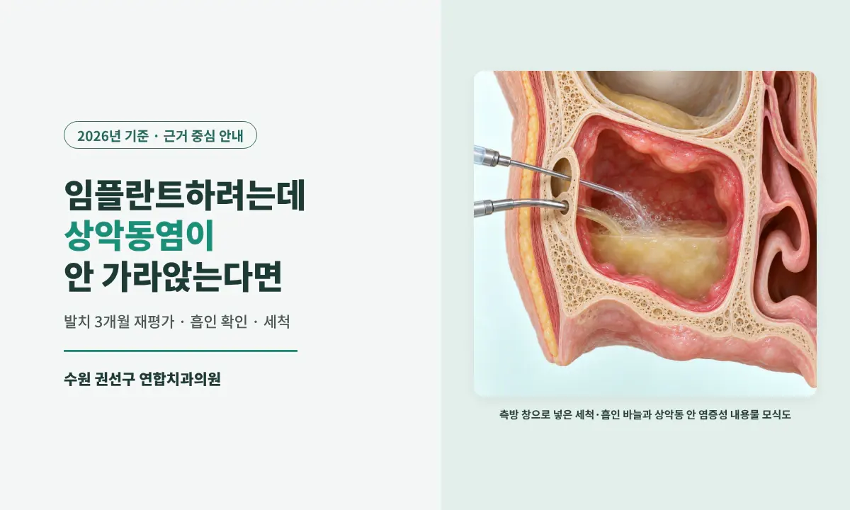
If sinusitis remains after an extraction and you are putting off your implant, what matters is less how long you wait than when it is rechecked and what the next step is. Three months after extraction, Yeonhap Dental Clinic uses CBCT to separate simple mucosal thickening from opacified lesions, and if the finding remains after one week of antibiotics, we open through a lateral approach and check directly.
Why doesn't my sinusitis settle after the tooth was extracted?
Some patients have had an upper molar extracted and, months later, are still putting off an implant because of sinusitis. The maxillary sinus is an air-filled space right above the roots of the upper molars, and sinusitis is inflammation inside it. When there is not enough bone in the upper molar area, a sinus lift is needed, in which the membrane on the floor of the sinus is raised and bone is filled in below it, but if inflammation remains, the order of this surgery changes.
[Key Answer]
Sinusitis that remains after extraction does not always resolve just by waiting, so it is important to set when it will be checked again and what the next step is. Yeonhap Dental Clinic looks again with a CBCT 3 months after extraction (a dental 3D CT scan) and checks for other tooth-related causes and for infection. If a cloudy finding remains inside the sinus even after treatment and we judge that direct treatment is needed, we check through a lateral approach (making a small window in the side wall of the sinus).
[4-Line Summary]
· Take a new CBCT 3 months after extraction, but simple mucosal thickening, where the lining looks only slightly thicker, is not by itself diagnosed as sinusitis and does not by itself delay an implant.
· If an opacified lesion, where the inside of the sinus looks cloudy and filled, remains, we check for other tooth-related causes and use antibiotics for 1 week.
· If it still remains, we open through a lateral approach and aspirate, and if inflammatory contents such as nasal-type discharge come out, we irrigate.
· After irrigation, if the remaining inflammation and the condition of the membrane allow us to proceed, we perform the sinus lift and bone graft, and if primary stability (the ability of the implant to stay firm without loosening right after placement) is actually achieved, we place the implant on the same day.
Written by: Dr. Ahn Seong-jin, Chief Director, Yeonhap Dental Clinic
[Table of Contents]
· Why doesn't my sinusitis settle after the tooth was extracted?
· When and based on what do we recheck sinusitis after extraction?
· If sinusitis remains after extraction, what comes first before an implant?
· Why don't we use the crestal approach for a sinus lift when sinusitis remains?
· What do we do when we open through a lateral approach because of sinusitis?
· When does ENT treatment come first when there is sinusitis?
When inflammation at the tip of an upper molar root or gum disease spreads to the maxillary sinus, it becomes odontogenic sinusitis (sinusitis that starts from a tooth). In the studies summarized in the Craig 2022 review, odontogenic sinusitis accounts for 25–40% of chronic sinusitis and 45–75% of cases in which only one maxillary sinus looks cloudy on CT.
Removing the causative tooth resolves most cases, but not all. The same review summarizes that even when the tooth is treated first, a considerable number of patients eventually needed sinus surgery. This happens when inflammation established inside the sinus continues even after the cause has been removed.
If a sinus lift is performed while inflammation remains, the membrane tears easily, and the bone graft material placed in an infected space can fail. That is why patients are told, "Come back once the inflammation has cleared," but if no one decides when to look again, the wait has no end. Yeonhap Dental Clinic sets the time to check and the next step for each result.
When and based on what do we recheck sinusitis after extraction?
Three months after extraction, we take a new CBCT and separate whether the lining looks only slightly thickened or the inside of the sinus looks cloudy and filled.
There are two reasons for using 3 months as the reference. It is roughly when the extraction site has healed enough to plan an implant, and inflammation that would resolve once the cause is removed shows change within this period. The reason we take a new scan rather than relying on the images from the time of extraction is that we need to see what has changed in between.
We do not diagnose sinusitis or postpone an implant just because the lining looks slightly thickened. If other lesions are seen together on CT, they are judged separately. The 2024 expert consensus by Lin et al. also states that mucosal thickening alone should not be diagnosed as odontogenic sinusitis. On the other hand, we do not pass over an opacified lesion (an area that looks blocked on CT) where the inside of the sinus looks cloudy and filled. It may be fluid that has collected, or thickened lining or a mass filling the space.
In this case, we first check whether there are other tooth-related causes, such as a neighboring tooth or a remaining root. If there are, we treat that tooth first, and if not, we regard it as a problem inside the sinus and move to the next step. If the finding has resolved, we decide whether a sinus lift is needed and which approach to use according to the remaining bone height. Those criteria are covered in Sinus Lift: Yeonhap Dental Clinic's 3 Treatment Criteria.
Course according to the sinusitis reassessment 3 months after extraction (Yeonhap Dental Clinic)
| CBCT finding | Judgment | Next step |
|---|---|---|
| No finding | Resolved after removing the cause | Plan sinus lift and placement according to remaining bone |
| Lining looks slightly thickened | Do not postpone for that reason alone | If other lesions are seen together, judge them separately |
| Opacified lesion + other tooth-related cause present | There is another cause | Treat that tooth, then reassess |
| Opacified lesion + no other cause | A problem inside the sinus | Antibiotics for 1 week |
| Opacified lesion after antibiotics | Do not conclude from imaging alone | Open through a lateral approach and confirm by aspiration |
If sinusitis remains after extraction, what comes first before an implant?
We use two antibiotics, amoxicillin and metronidazole, for 1 week and see how you respond.
Bacteria in the mouth, particularly anaerobic bacteria that grow without oxygen, are involved in odontogenic sinusitis, so this combination targets both. The 2024 consensus by Lin et al. also lists antibiotics as an important option for odontogenic sinusitis with clear bacterial infection.
However, antibiotics alone do not always finish the job. The Craig 2022 review summarizes that oral antibiotics alone usually do not resolve odontogenic sinusitis. So at our clinic, the 1 week of antibiotics is a step to identify cases that settle with medication. If it improves, we move on to the sinus lift and placement as planned, and we do not add surgery where medication is enough.
If there is no improvement, we do not keep waiting. If we have waited 3 months, there is no other cause, and there is no response to antibiotics, we judge that it is time to look inside the sinus directly.
Why don't we use the crestal approach for a sinus lift when sinusitis remains?
The sinus lining that has stayed inflamed is swollen and weak, so it tears easily while being pushed up from below, and we also cannot check the inside directly.
The crestal approach to a sinus lift raises the membrane from below through the small hole where the implant will go. When the membrane is healthy, Yeonhap Dental Clinic considers this approach first, especially the hydraulic lift that raises the membrane with saline pressure. It is done without an incision or with a minimal incision, so swelling tends to be less.
We do not use this approach when sinusitis remains. First, the inflamed lining is swollen and weak, so a perforation (a tear or hole in the sinus membrane) is likely to occur while lifting. Second, because the procedure goes through a small hole, we can neither check whether the inside of the sinus holds pus or clear fluid nor wash it out. What is needed now is not raising the membrane but directly checking and treating the inside of the sinus.
How perforation is checked in the crestal approach and when we switch to the lateral approach is covered separately in Limits of the Crestal Sinus Lift Approach and How to Overcome Them.
Even a familiar method is taken off the list when the conditions do not fit. We believe the approach is determined by the condition of the sinus, not by what is comfortable for the operator.
Sinus lift approach according to sinus condition (Yeonhap Dental Clinic)
| Sinus condition | Approach | Reason |
|---|---|---|
| Healthy membrane (including a lining that looks only slightly thickened) | Crestal approach (hydraulic lift) first | No incision or a small one, so less swelling |
| Opacified lesion that improves after antibiotics | Consider the crestal approach | Decide after rechecking the membrane condition |
| Opacified lesion that remains after antibiotics | Lateral approach | Need to see directly, aspirate, and irrigate |
| Clear fluid found on opening laterally | Usually lift and bone graft without irrigation | Decide by looking together at the contents, membrane condition, and remaining inflammation |
| Aspiration is poor and a mass is found (fungal ball suspected) | Remove the mass; the lift is usually done at a later visit | The membrane is often significantly damaged during removal |
What do we do when we open through a lateral approach because of sinusitis?
We make a window in the side wall of the sinus, aspirate to check the contents, and irrigate if needed. If the remaining inflammation and the membrane condition allow us to proceed, we perform the sinus lift and bone graft, and if primary stability is actually achieved, we place the implant on the same day.
The lateral approach makes a small window in the side wall of the sinus (the bone on the cheek side) and proceeds while looking directly at the membrane. It involves a larger incision and area than the crestal approach, but in this situation the ability to check and treat the inside directly matters more.
The sequence is as follows.
· Make a window in the side wall, insert a needle past the membrane, and aspirate the contents of the sinus
· If inflammatory contents such as nasal-type discharge come out, wash them out with saline and chlorhexidine disinfectant while aspirating at the same time (irrigation)
· If clear fluid comes out, we look together at the contents, membrane condition, and remaining inflammation, and at our clinic we usually move on to the lift without irrigation
· The small holes left by the irrigation and aspiration needles are closed with a resorbable barrier membrane (a thin membrane that the body absorbs over time)
· If the remaining inflammation and the membrane condition allow us to proceed, we raise the membrane and fill in bone graft material
· If at least 1 mm of bone remains and primary stability is actually secured, we place the implant on the same day
The next step depends on the aspiration result because imaging alone cannot tell them apart. A cloudy finding on CT looks similar whether the sinus holds pus, clear fluid, thickened lining, or a mass. That is why we do not conclude sinusitis from imaging alone, and decide whether to irrigate by looking at the aspiration result together with the membrane condition.
If inflammatory contents keep coming out after irrigation, or the membrane is torn so extensively that it cannot be repaired, we only irrigate and postpone the lift. This is uncommon but can happen. Even after the lift and bone graft are done, if primary stability is not achieved we do not place the implant on the same day; we wait for the bone to settle and place it, usually 4–5 months later.
Sometimes aspiration is not possible at all. If the sinus looks densely cloudy on CT and denser, millet-sized spots are seen here and there, a fungal ball (a clump of fungus) is suspected. Cha et al. 2020 analyzed 247 people who had endoscopic surgery for one-sided sinusitis and reported that dense spots (calcification) within the lesion were the CT finding most commonly associated with a fungal ball, while also concluding that diagnostic accuracy improves when other features are looked at together. Dense spots or poor aspiration are clues for suspicion, and confirming the diagnosis requires pathology examination of the removed tissue.
At our clinic, when aspiration is poor we look directly inside, and if a mass that needs removal is found we remove it with instruments. In this process the sinus membrane is often significantly damaged, so in my practice I did not do the lift on the same day in such cases and postponed it until the membrane had recovered. The sequence for handling sinus lesions, including fungal balls, is also summarized in Complex Implants: Four Things We Check Before Placement.
There are also reports on irrigation treatment itself from Korea. Jaiswal et al. 2024 irrigated with saline or iodine solution through a small lateral window in 21 patients with odontogenic sinusitis (most of whom developed it after dental procedures such as implants, bone grafts, or extractions) and reported that the volume of cloudy area on CBCT decreased by 40.15% and symptoms improved in 17 of the 21 (80.9%). It was a retrospective study of irrigation alone, not one that combined a same-day lift and placement. Same-day lift and placement is our clinic's selection criterion.
Gang et al. 2021 reported that, in 40 patients who had surgery for a large cyst involving the maxillary sinus, the group irrigated with saline after surgery had significantly less facial swelling, pain, and fever than with the traditional Caldwell-Luc procedure (a traditional operation that opens the front wall of the sinus and removes the diseased lining). Note that this was a study of cyst surgery patients, not sinusitis.
When does ENT treatment come first when there is sinusitis?
ENT treatment comes first when the inflammation has spread to other sinuses, or when the passage through which the maxillary sinus drains into the nose itself needs to be opened.
The dental clinic takes on this process because the cause started in a tooth and the plan must eventually lead to an implant. The Craig 2022 review also states that, as a rule, odontogenic sinusitis is managed jointly by ENT and dentistry. In the following situations, ENT treatment comes first.
· When the inflammation has spread to other sinuses such as the ethmoid sinus (the sinus on the inner side between the eyes) — Crovetto-Martínez et al. 2014 reported that, among 55 patients with odontogenic sinusitis who had endoscopic sinus surgery, 52.7% had spread to the anterior ethmoid sinus (a retrospective study looking back at patients who went on to surgery)
· When complications spreading around the eyes or toward the head are suspected
· When the natural ostium (the opening through which the maxillary sinus drains into the nose) is blocked and the drainage pathway itself needs to be opened
· When inflammation recurs even after irrigation
5 things to check when consulting about an implant with sinusitis in Gwonseon-gu and Tap-dong, Suwon
The practical standard for a consultation is whether the waiting period has an end.
Yeonhap Dental Clinic, across from Suwon NongHyup Guun Branch (231 Geumho-ro), on the 5th floor at 230 Geumho-ro, Gwonseon-gu, Suwon, Gyeonggi-do (Tap-dong, Cham Clinic Building), has treated patients in the same place since 2002, and Dr. Ahn Seong-jin, Chief Director and Integrated Dentistry specialist, oversees treatment.
· Whether a time to check again has been set
· Whether a new CBCT is taken then to compare the change
· Whether they explain separately a slightly thickened lining and a sinus that looks cloudy and filled
· Whether they also check if other tooth-related causes remain
· Whether they explain the conditions for doing the lift and placement on the same day or splitting them
[Key Takeaways]
✓ Sinusitis can remain even after the causative tooth is extracted, so Yeonhap Dental Clinic takes a new CBCT 3 months after extraction to separate simple thickening from opacified lesions.
✓ If an opacified lesion remains and there is no other tooth-related cause, we use antibiotics for 1 week, and if it still remains, we open through a lateral approach instead of the crestal approach and confirm by aspiration.
✓ After irrigation, if the remaining inflammation and the membrane condition allow us to proceed, we perform the lift and bone graft, and if primary stability is actually secured, we place the implant on the same day.
A Few Words from the Treatment Room About Sinusitis and Implants
In the treatment room I often hear, "I thought it would clear up once the tooth was out, but it's been the same for months." First, on the new CBCT taken 3 months later, I separate whether the lining looks only slightly thickened or the inside of the sinus looks cloudy and filled. I do not postpone an implant just because the lining looks slightly thickened; if it is cloudy and filled, I try antibiotics, and if it still remains, I open from the side and check directly. When the membrane is healthy, I try to lift it from below without an incision and place the implant on the same day even if 1 mm or more of bone remains, but I leave that method out at a site where sinusitis remains. In my practice, I opened, aspirated, and irrigated if it was inflammatory, and if it was clear fluid and the membrane condition was fine, I lifted as it was, and in most cases I finished the lift, bone graft, and placement on the same day. So far I have not had a case where the membrane was damaged after washing out pus and I had to postpone the lift, but in a case where aspiration was poor, I looked inside and removed a mass that looked like a fungal ball, and the membrane was significantly damaged in that process, so I postponed the lift to a later visit. This is why, even when the explanation is the same "you have sinusitis," the sequence differs depending on the condition inside the sinus.
References
📋 Treatment Information
This post was written in compliance with the Korean Medical Service Act and the Ministry of Health and Welfare's medical advertising guidelines, and is intended to provide medical information. It does not guarantee or assert the result of any specific treatment. The treatment that can be applied and the course of treatment differ from person to person depending on the condition of the sinus and the remaining bone, so please be sure to consult fully with your dental team before deciding on diagnosis and treatment direction.
🦷 Sinus lift information: Sinus Lift Implants in Suwon
📍 5th Floor, 230 Geumho-ro, Gwonseon-gu, Suwon, Gyeonggi-do (Tap-dong, Cham Clinic Building)
🚕 Across from Suwon NongHyup Guun Branch (231 Geumho-ro) / about 15 minutes by taxi from Suwon Station
🚗 Free parking available in the building
📞 031-293-2207
🌐 yeonhapdental.com
🟡 Opened in 2002 / Treatment overseen by Dr. Ahn Seong-jin, Chief Director and Integrated Dentistry specialist
🔗 Online booking and consultation
Sinusitis and Implants: Frequently Asked Questions
- What should I do if sinusitis doesn't go away after the tooth is extracted?
- Even after the causative tooth is extracted, inflammation inside the maxillary sinus (the air-filled space above the upper molars) can continue. Yeonhap Dental Clinic takes a new CBCT (a dental 3D CT scan) 3 months after extraction to check, and if it remains, we first look for other tooth-related causes.
- If the CT says my sinus lining is thick, can't I have an implant?
- We do not diagnose sinusitis or postpone an implant just because the lining looks slightly thickened. If other lesions are seen together on CT, they are judged separately. The expert consensus (Lin et al. 2024) is that thickening of the lining alone is not diagnosed as sinusitis, and we move to the next step when the inside of the sinus looks cloudy and filled.
- How much longer do I have to wait for an implant because of sinusitis?
- We do not leave the waiting period open-ended. We reassess 3 months after extraction, and if the inside of the sinus looks cloudy and filled, we use antibiotics for 1 week, and if it still remains, we open through a lateral approach (making a window in the side wall of the sinus) to check and treat.
- Even with sinusitis, can't it be done with a method that needs no incision?
- Not in this situation. The crestal approach, which raises the membrane from below without an incision, does not let us see inside the sinus, and a membrane that is swollen and weakened by inflammation is likely to perforate (tear or develop a hole) when lifted. It is a method used when the membrane is healthy.
- What if I open the sinus and it isn't pus?
- If clear fluid comes out, we look together at the contents, membrane condition, and remaining inflammation, and at our clinic we usually move on to the sinus lift without irrigation. On CT, pus, clear fluid, and thickened lining can look similar, so we do not conclude until we have opened and checked.
- Do I have to come back for the implant a few months after sinus irrigation?
- If conditions allow, we do it on the same day. After irrigation, if the remaining inflammation and the membrane condition allow us to proceed, we perform the sinus lift (surgery that raises the membrane and fills in bone) and bone graft, and if primary stability (the ability of the implant to stay firm without loosening right after placement) is actually secured, we place the implant on the same day. If primary stability is not achieved, we usually place it 4–5 months later.
- Does the lateral approach to the sinus cause a lot of swelling?
- The incision area is larger than with the crestal approach, so there tends to be more swelling. In return, checking, irrigating, and lifting can be done in one session while looking directly at the membrane, which can reduce the burden of splitting the surgery into several stages.
- When do I need to go to an ENT for sinusitis?
- These are cases where the inflammation has spread to other sinuses such as the ethmoid sinus, where complications around the eyes or toward the head are suspected, or where the passage through which the maxillary sinus drains into the nose itself needs to be opened. If it recurs after irrigation, we consider it together.
Related pages
Before your visit
This article provides general information on oral health and dental care. Whether treatment is needed, and which method is appropriate, may change after an oral exam and any necessary diagnostic tests. If you have pain, a loose tooth, swelling, or discomfort with a prosthesis, please visit us for a consultation.