
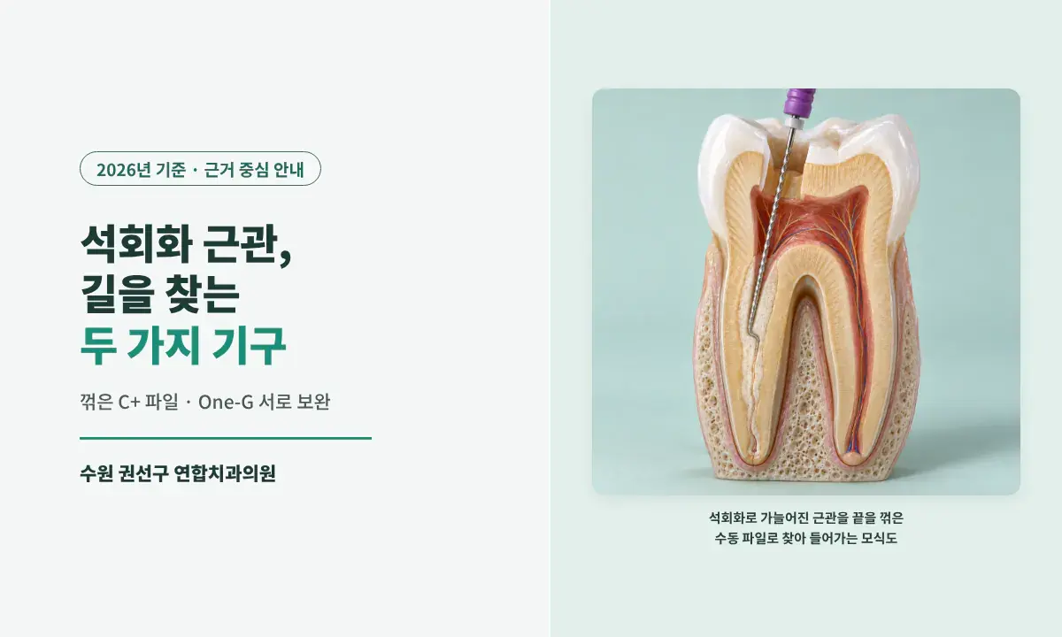
A calcified root canal does not mean root canal treatment starts right away. Yeonhap Dental Clinic in Gwonseon-gu, Suwon, decides whether treatment is needed by looking at symptoms, pulp testing, and root-tip imaging together; for calcified canals that do need treatment, we find the path with a pre-curved C+ file and an One-G glide path file, which complement each other depending on where and how the canal is blocked.
What is canal calcification, and does a calcified canal always need root canal treatment?
If you are told at a check-up that "the canal is blocked," it is easy to worry that the tooth is hard to treat. Inside a tooth root is a thin channel called the root canal, through which the nerve and blood vessels run, and root canal treatment removes infected nerve tissue from this canal, disinfects it, and seals it. Canal calcification means hard tissue has built up inside this channel so that the passage has narrowed or closed.
[Key Answer]
Canal calcification (canal obliteration) is not in itself something to treat; whether root canal treatment is needed is judged by looking at symptoms, pulp testing, and the root-tip imaging findings together. In calcified canals that need root canal treatment, when a #10 K-file (a thin stainless steel hand file) does not advance well, Yeonhap Dental Clinic finds the path in most cases with a C+ hand file with a bent tip, and for canals blocked right from the entrance we also use an One-G glide path file (a nickel-titanium rotary instrument that prepares a passage for the rotary files to follow).
[Summary in 4 Lines]
· We do not start root canal treatment just because a canal is calcified; we decide by looking at symptoms, pulp testing, and root-tip imaging together.
· To find the path, we use a C+ file with a bent tip and One-G as complements depending on where and how the canal is blocked, and One-G is at a disadvantage in bent paths such as a ledge (a step formed on the canal wall).
· In laboratory studies, manual K-files caused more canal transportation (deviation from the original path) than machine-driven files, but there is no evidence that One-G is better than other rotary files (Aminsobhani 2022, Shojaeian 2021).
· Regardless of how far the canal is opened, we keep sodium hypochlorite (NaOCl) of 6% or higher in contact for a cumulative 20 to 40 minutes, and if the canal never opens, we divide the plan according to the root-tip lesion and whether the tooth can be saved.
Written by Dr. Ahn Seong-jin, Chief Director, Yeonhap Dental Clinic
[Table of Contents]
· What is canal calcification, and does a calcified canal always need root canal treatment?
· In what order do you find the path in a calcified canal?
· When is the One-G file used in a calcified canal, and when is it not?
· How is disinfection done in a calcified canal?
· What happens if a calcified canal never opens?
· How is a tooth managed after root canal treatment of a calcified canal?
The root canal does not stay the same width throughout life. With age, trauma, or ongoing irritation under an old restoration, hard tissue gradually builds up on the canal wall. A review summarizes studies reporting canal obliteration following trauma, orthodontics, and replantation (putting a lost tooth back in place) at 3.8 to 24% (Chaniotis 2024).
Calcification usually causes no symptoms and is found by chance on check-up images. We do not start root canal treatment simply because a canal is calcified. We decide whether treatment is needed by looking at symptoms, pulp testing (a test of whether the nerve is alive), and root-tip imaging findings together.
A literature review pooling cases of front teeth with calcified canals after trauma reported cases in which some teeth without a root-tip lesion were simply monitored without treatment. Because this pools case reports, however, it cannot be taken as a figure representing the prognosis of all calcified teeth (Vinagre 2021).
Difficulty arises when root canal treatment becomes necessary because of pain or a root-tip lesion. If the passage is narrow or blocked, instruments struggle to reach the space that needs disinfection, and forcing them can deviate from the original path, creating a ledge in the canal wall or a perforation, a hole through the root. That is why, in a calcified canal, which instrument is used in what order matters.
In what order do you find the path in a calcified canal?
After locating the canal orifice, we start with a #10 K-file, and if it does not catch well, we find the path with a pre-curved C+ file.
If the canal is hard to see on a periapical radiograph (a small X-ray that captures the root tip), we expect calcification and prepare for it. If we need to see the canal shape better, we add a 3D CT, but we do not take one in every case.
When we open the chewing surface and enter with a #330 bur (a small rotary instrument for opening the tooth), there is usually a sensation of the instrument suddenly dropping in (a drop-in) as it reaches the empty space where the nerve was (the pulp chamber). When a drop-in is felt, we clean up the orifice with an Endo-Z bur and locate the canal orifice with an endodontic explorer (a sharp-tipped probe).
With heavy calcification, this space is filled with hard tissue and no drop-in is felt. At Yeonhap Dental Clinic, when there is no drop-in, or the canal orifice is still not visible after opening to the expected depth, we then take a periapical radiograph or CT to check the depth and direction. This is because continuing to cut without that sensation can create a perforation, a hole through the side of the root.
For an uncomplicated canal, we confirm patency (the state in which a file passes through to the canal's end opening) with a #10 K-file and then enlarge it with nickel-titanium rotary files. If a K-file does not catch well, we bend the last 1 mm of a #10 C+ file to 90° and use it. By turning the tip direction, the file feels its way through narrow orifices and bent passages, and in my practice, most canals where a K-file does not catch well open at this stage.
In an experiment on narrow canals, the manual K-file group showed more canal transportation near the root tip than the OGP reciprocating file group, and six #10 K-files were severely deformed in the manual group. However, this was not a study comparing One-G, so it cannot be used as evidence of One-G's superiority (Liu 2021).
In an experiment with 100 S-shaped resin practice blocks, the #15 K-file group showed more canal transportation at the root tip and middle than the nickel-titanium rotary file groups including One-G (p<0.001), with no significant difference among the rotary file groups. This was a resin block experiment, not real teeth, and all groups were compared after first creating a path with a #10 manual file (Aminsobhani 2022).
When is the One-G file used in a calcified canal, and when is it not?
One-G helps in canals blocked right from the entrance, but it is at a disadvantage in bent paths such as a ledge, where we return to the pre-curved C+ file.
One-G is a nickel-titanium rotary instrument that prepares a glide path (a passage for rotary files to follow), and its manufacturer lists narrow or heavily calcified canals and highly curved canals as indications. It is more flexible than a stainless steel hand file and advances little by little as it rotates without applying force.
When using it, we do not push it in; we wait for the sensation of the instrument boring in on its own, and withdraw it right away when we feel the file binding on the wall, repeating this. We also check from time to time whether the file blades are damaged. Continuing with a damaged file can throw off the direction or cause it to break.
A ledge is a step-shaped shelf on the canal wall; it sometimes results from instruments being forced in earlier treatment, and a calcified canal can itself have such a bent shape. A rotary instrument tends to bore toward the side of least resistance, and when there is a ledge, that direction is toward the ledge rather than the original path. Continuing to use it deepens the ledge and raises the risk of perforation, so in this case we use the pre-curved C+ file to ride over the ledge and find the original path.
C+ and One-G are not instruments to be swapped in a fixed order; they complement each other depending on where and how the canal is blocked.
A study comparing 30 curved, healthy canals with micro-CT found no significant difference in canal transportation or centering ability between One-G and two other rotary files. It was not a study of calcified canals, and even taken together with the resin block experiment above, it is not evidence that One-G is better than other rotary files (Shojaeian 2021).
Choosing path-finding instruments for calcified canals (Yeonhap Dental Clinic)
| Canal situation | Instrument used at Yeonhap Dental Clinic | Reason |
|---|---|---|
| Uncomplicated canal | Check patency with a #10 K-file, then nickel-titanium rotary files | First confirm that the path is open to the end of the canal |
| Canal where a K-file does not catch well | #10 C+ file with the last 1 mm bent 90° | Change the tip direction to find the narrow orifice and bent passage |
| Canal blocked right from the entrance | One-G glide path file | Advances little by little by rotation without applying force |
| Bent path such as a ledge | Back to the pre-curved C+ file | A rotary instrument bores toward the ledge, raising the perforation risk |
How is disinfection done in a calcified canal?
Regardless of how far the instrument reaches, we keep to the same disinfection sequence: placing a rubber dam and keeping NaOCl of 6% or higher in contact for a cumulative 20 to 40 minutes.
In an experiment that examined extracted upper molars with micro-CT, 35% or more of the canal surface remained unchanged before and after shaping with all four nickel-titanium shaping techniques (Peters 2001). This is the result of that experiment and does not mean the same proportion applies to every treatment, but it shows that some surfaces instruments cannot reach may remain, so the role of the irrigant is large.
· Rubber dam (a rubber sheet that covers the mouth, leaving only the tooth being treated exposed): used in every root canal treatment, and for a tooth with a collapsed wall, we build up the wall first before placing it.
· NaOCl: we use a solution of 6% or higher concentration as labeled, undiluted. From the time the canal is enlarged to the planned size until obturation (sealing the disinfected canal with filling material), we replace it every 5 minutes, and the soaking time across visits is added up to give a cumulative 20 to 40 minutes of contact.
· EDTA: in heavily calcified canals, we rinse with EDTA (an irrigant that helps remove the mineral component and smear layer of the canal) and then finish with NaOCl.
· Calcipex (calcium hydroxide medicament): placed inside the canal between visits; the interval is usually 1 week and does not exceed 1 month.
When the cumulative soaking time reaches 20 minutes, we check whether exudate (fluid seeping from inside the canal) or bubbles keep rising, together with the condition of the canal, to decide whether to fill. If they continue, we soak up to a cumulative 40 minutes, and if they persist even after that, we enlarge and irrigate further that day, place Calcipex, and reassess at the next visit. Exudate or bubbles alone do not establish that infection remains or that the enlargement is insufficient; this is our clinic's way of proceeding.
The number of visits is usually 2 for front teeth and premolars and 3 for molars, and 1 to 2 more may be needed if calcification is heavy or the root-tip lesion is large. The rubber dam and disinfection sequence are covered in detail in Why a Rubber Dam Matters in Root Canal Treatment.
What happens if a calcified canal never opens?
We do not dig further; after disinfecting as far as we can reach, we divide the plan according to why treatment was needed in the first place, the root-tip lesion, and whether the tooth can be saved.
There are canals that do not open all the way even when the C+ file and One-G are used together. Digging deeper then raises the risk of perforation, so Yeonhap Dental Clinic disinfects and fills as far as the instruments reach and tells you plainly that there is a section that did not open. This is because you need to know from the start, so that if a problem arises later, we can judge the cause together.
We view a case with no root-tip lesion but a section that did not open differently from a case with a lesion and a section that did not open. A lesion suggests that bacteria are likely to remain in the canal, and a section the instruments did not reach is hard to disinfect further from inside. In that case, we consider an apicoectomy (root-end resection), which removes the root tip and surrounding inflammation from the gum side after finishing with a crown, and Yeonhap Dental Clinic performs apicoectomy itself.
If the periodontal condition (the state of the gums and the bone supporting the teeth) is poor and the tooth is hard to save even with apicoectomy, we recommend extraction and discuss separately how to replace the tooth afterward. If extraction is not needed right away but the long-term prognosis is uncertain and you do not want an implant, we explain the limits and then restore the tooth with a resin core, without a crown, and monitor it. Even for a tooth we have recommended extracting, the final decision is yours, so we explain thoroughly what problems may arise if it is not extracted.
Monitoring a tooth that has only calcification, with no symptoms or lesion, so root canal treatment was not started, is not the same observation as monitoring a canal that did not open after treatment was started because of infection. The table below separates the two.
What we do when a calcified canal never opens (Yeonhap Dental Clinic)
| Situation | Yeonhap Dental Clinic's approach | Next options |
|---|---|---|
| Calcification only, no symptoms or lesion | Root canal treatment not started; checked together at check-ups | Consider root canal treatment if symptoms or a root-tip lesion appear |
| Part of the canal not opened during treatment, no lesion | Disinfect and fill as far as reachable, inform the patient, then core and crown | Consider apicoectomy if a lesion or symptoms appear during follow-up |
| Not opened, with a root-tip lesion | Disinfect and fill as far as reachable, consider apicoectomy after the crown | Consider extraction and an implant if the periodontal condition is poor and the tooth is hard to save |
| Hard to save | Judge the need and timing of extraction | Discuss separately how to replace the tooth after extraction |
How is a tooth managed after root canal treatment of a calcified canal?
Every 6 months we check symptoms and radiographs to see whether the lesion is shrinking, and if it grows or symptoms appear, we consider the next step.
A root-tip lesion takes time to heal, so we do not judge treatment a failure simply because one remains right after treatment. Yeonhap Dental Clinic checks symptoms and radiographs every 6 months to see whether the lesion is shrinking. If the lesion grows or pain, swelling, or a sinus tract (a pus drainage hole in the gum) appears, we consider additional treatment, and even without symptoms, if it does not shrink over a long period, we reassess the cause and the need for treatment.
Apicoectomy is surgery that cuts off the root tip, so it reduces the part of the tooth that supports it by that much. That is why we do not rush into it for a stable tooth.
5 things to check when consulting about root canal treatment for a calcified canal in Gwonseon-gu or Tap-dong, Suwon
If you have been told your canal is blocked, the practical test of a consultation is whether the dentist explains which instruments and which order they will use depending on the shape of the blockage.
Yeonhap Dental Clinic, at 230 Geumho-ro, 5th Floor (Cham Clinic Building, Tap-dong), Gwonseon-gu, Suwon, across from Suwon NongHyup Guun Branch (231 Geumho-ro), has been treating patients in the same location since 2002, with Dr. Ahn Seong-jin, Chief Director and Integrated Dentistry specialist, in charge. You can see the process in Root Canal and Conservative Treatment Information.
· Whether treatment is recommended for calcification alone, or decided by looking at symptoms, pulp testing, and imaging together
· Which instruments are used in which order when the canal does not open easily
· Whether depth is checked with a periapical radiograph or CT when the canal orifice cannot be found
· Whether you are told in advance how the case will be finished, depending on the root-tip lesion, if the canal never opens
· Whether conservative options such as apicoectomy are considered before extraction
[Key Takeaways]
✓ Canal calcification is not in itself something to treat; Yeonhap Dental Clinic decides on root canal treatment by looking at symptoms, pulp testing, and root-tip imaging together.
✓ If a K-file does not catch well, we find the path in most cases with a pre-curved C+ file, use One-G in canals blocked from the entrance, and go back to C+ in bent paths such as a ledge. There is no evidence that One-G is better than other rotary files.
✓ If the canal never opens, we disinfect as far as we can reach and tell you so, then consider a crown, apicoectomy, and extraction in that order depending on the root-tip lesion and whether the tooth can be saved.
What I would like to tell you about root canal treatment of calcified canals from the treatment room
When I see a calcified tooth, I first ask whether it truly needs root canal treatment. A canal that merely looks blocked on an image is not a reason to treat; I start only after looking at symptoms, pulp testing, and the condition of the root tip together. Once treatment begins, I first feel the path with a #10 K-file, and if it does not catch well, I bend the tip of a C+ file and go in, and in my practice most canals open at this point. If the canal is blocked right from the entrance, One-G helps, but in a bent path such as a ledge it is a disadvantage, so I return to the bent C+. No single instrument is a cure-all; I think the key is swapping the two instruments to suit the shape of the blockage. Still, some canals never open all the way, and in that case I do not dig further: I disinfect as far as I can reach, tell you plainly, and if there is a root-tip lesion, discuss options up to apicoectomy together with you.
References
📋 Clinic Information
This post was written in accordance with the Korean Medical Service Act and the Ministry of Health and Welfare's medical advertising guidelines, and is intended to provide medical information. It does not guarantee or assert the result of any particular instrument or treatment. The applicable methods and course differ from person to person depending on the canal condition and root-tip conditions, so please decide on a diagnosis and treatment direction only after a thorough consultation with your dental team.
🦷 Root canal and conservative treatment information: Yeonhap Dental Clinic root canal treatment
📍 230 Geumho-ro, 5th Floor (Cham Clinic Building, Tap-dong), Gwonseon-gu, Suwon, Gyeonggi-do
🚕 Across from Suwon NongHyup Guun Branch (231 Geumho-ro) / about 15 minutes by taxi from Suwon Station
🚗 Free parking available in the building
📞 031-293-2207
🌐 yeonhapdental.com
🟡 Open since 2002 / Treatment overseen by Dr. Ahn Seong-jin, Chief Director and Integrated Dentistry specialist
🔗 Online booking and consultation
Frequently asked questions about root canal treatment of calcified canals
- Does a calcified canal always need root canal treatment?
- No. Canal calcification (a state in which hard tissue has built up inside the root canal so that the passage has narrowed or closed) is not in itself something to treat. Yeonhap Dental Clinic decides whether root canal treatment is needed by looking at symptoms, pulp testing (a test of whether the nerve is alive), and root-tip imaging findings together.
- What instruments are used to open a calcified canal?
- Yeonhap Dental Clinic starts with a #10 K-file (a thin stainless steel hand file), and if it does not catch well, we find the path in most cases with a C+ file whose last 1 mm is bent 90°. For canals blocked right from the entrance, an One-G glide path file (a rotary instrument that prepares a passage for rotary files to follow) helps, and we use the two instruments as complements depending on where and how the canal is blocked.
- Is the One-G file better than other glide path files?
- There is no evidence to say so. In a micro-CT comparison of curved canals, there was no significant difference in canal transportation (deviation from the original path) or centering ability between One-G and other rotary files (Shojaeian 2021), and in a resin block experiment, there was no difference among the rotary files either (Aminsobhani 2022).
- Is the One-G file used in a calcified canal with a ledge?
- In bent paths such as a ledge (a step-shaped shelf on the canal wall), One-G is at a disadvantage. A rotary instrument tends to bore toward the ledge, the side of least resistance, raising the perforation risk, so in this case Yeonhap Dental Clinic uses a pre-curved C+ file to ride over the ledge and find the original path.
- How many visits does root canal treatment of a calcified canal take?
- At Yeonhap Dental Clinic, treatment is usually completed in 2 visits for front teeth and premolars and 3 for molars, and 1 to 2 more may be needed if calcification is heavy or the root-tip lesion is large. Between visits we place Calcipex (calcium hydroxide medicament); the interval is usually 1 week and does not exceed 1 month.
- If a calcified canal never opens, do I have to have the tooth extracted?
- We do not extract right away. Yeonhap Dental Clinic disinfects and fills as far as we can reach and tells you there is a section that did not open, and if there is a root-tip lesion, we consider an apicoectomy (surgery that removes the root tip and inflammation from the gum side) after the crown. When the periodontal condition is poor and the tooth is hard to save, we recommend extraction and discuss separately how to replace the tooth afterward.
- If a lesion remains after root canal treatment of a calcified canal, do I have to be treated again?
- Not necessarily right away. Yeonhap Dental Clinic checks symptoms and radiographs every 6 months to see whether the root-tip lesion is shrinking. If the lesion grows or pain, swelling, or a sinus tract (a pus drainage hole in the gum) appears, we consider additional treatment, and even without symptoms, if it does not shrink over a long period, we reassess the cause and the need for treatment.
- Where is Yeonhap Dental Clinic in Tap-dong and Guun-dong, Gwonseon-gu, Suwon?
- We are at 230 Geumho-ro, 5th Floor (Cham Clinic Building, Tap-dong), Gwonseon-gu, Suwon, Gyeonggi-do, across from Suwon NongHyup Guun Branch (231 Geumho-ro). Free parking is available in the building, and we are about 15 minutes by taxi from Suwon Station.
Related pages
Before your visit
This article provides general information on oral health and dental care. Whether treatment is needed, and which method is appropriate, may change after an oral exam and any necessary diagnostic tests. If you have pain, a loose tooth, swelling, or discomfort with a prosthesis, please visit us for a consultation.