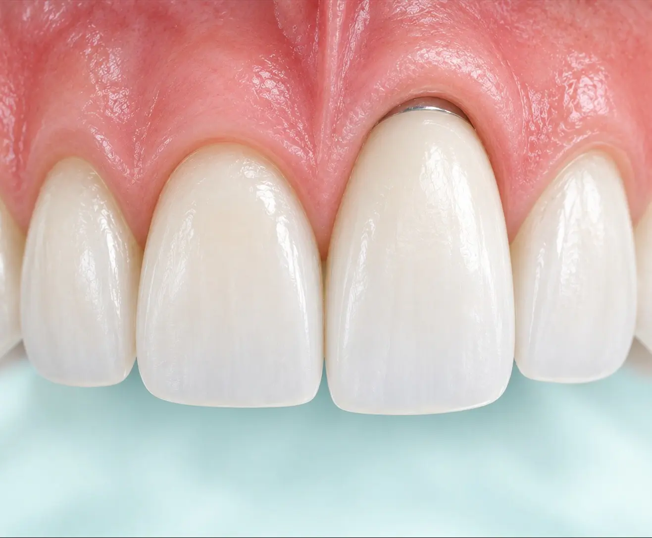
Front-view diagram comparing front tooth #11 placed in its proper position with implant tooth #21 placed too far outward, where the gum has risen and the tooth looks long

FRONT TOOTH IMPLANT

Front Tooth Implants Three Criteria for Placing Them Properly

Position, angle, and bone: when all three are right, the gum line matches the neighboring teeth. So if conditions are met, we place it on the day of extraction, toward the palate, at an angle within the incisal edge line.

Why Diagnosis Matters

A front tooth implant is a tug-of-war between “bone” and “prosthesis length”

Front teeth have thin cheek-side bone and are visible the moment you smile, so 2 mm of bone and the gum line together decide the result. So the implant is placed close to the palate to secure the cheek-side bone, and the axis is tilted toward the cheek by the same amount to match the direction of the prosthesis. If the axis crosses the line connecting the incisal edges of the neighboring teeth, the crown becomes long, so the tilt goes only as far as that line. The cheek-side gap is filled with a bone graft to make 2 mm, and if infection can be removed, the gum has not collapsed, and initial stability is achieved, it is placed on the day of extraction.

Consultation Checklist

What we check at your first consultation

At the first consultation, we check the five items below using 3D CT and your smile.

  • How much gum shows when you smile. For front teeth, the gum line is the result
  • Cheek-side bone thickness: whether a diameter that still leaves 2 mm after placing the implant is available
  • Whether, when placed toward the palate with the axis tilted toward the cheek, an angle that does not cross the neighboring incisal edge line is achievable
  • If the tooth needs to be extracted, whether infection can be removed and whether the gum has collapsed
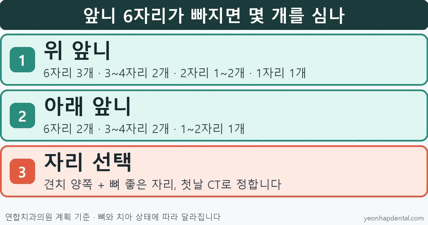
  • The number of missing sites. The standard is 3 for the six upper front tooth sites and 2 for the six lower sites

Treatment Options

Three ways front tooth implants differ from molars

Position and angle, the result, and placement on the day of extraction.

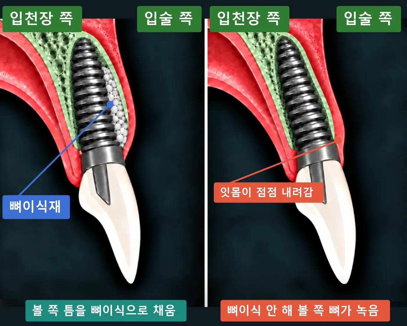
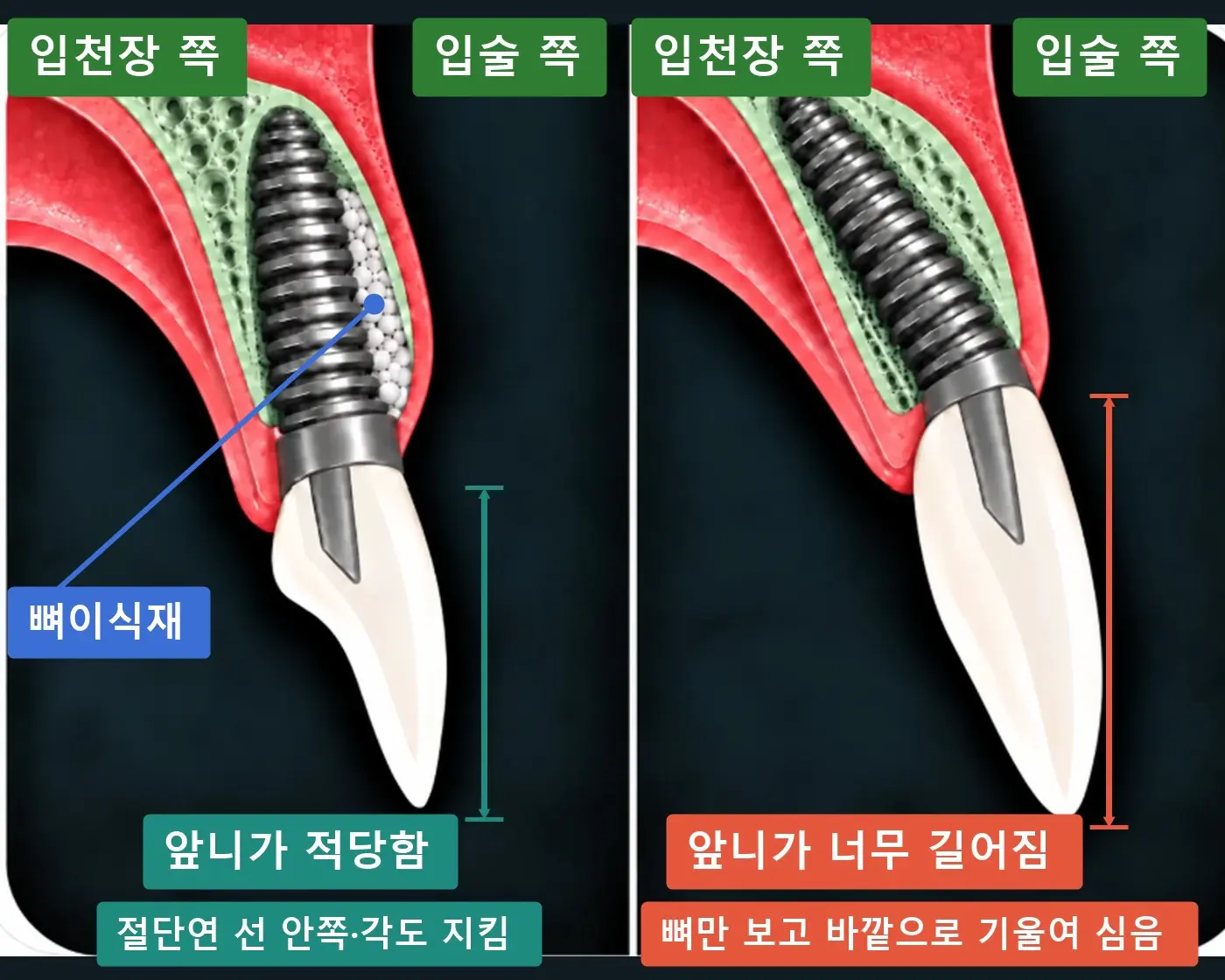
Mediolateral cross-section diagram comparing a front tooth implant placed within the incisal edge line with one tilted toward the cheek past the line, resulting in a longer crown

Place it toward the palate, and tilt the axis toward the cheek only within the incisal edge line

To secure the cheek-side bone, the implant is placed close to the palate, and the axis is tilted toward the cheek by the same amount to match the direction of the prosthesis. At Yeonhap Dental Clinic, the limit of that tilt is the line connecting the incisal edges of the neighboring teeth. If the axis crosses this line, it is a danger zone where the crown becomes long, so we do not place at that angle.

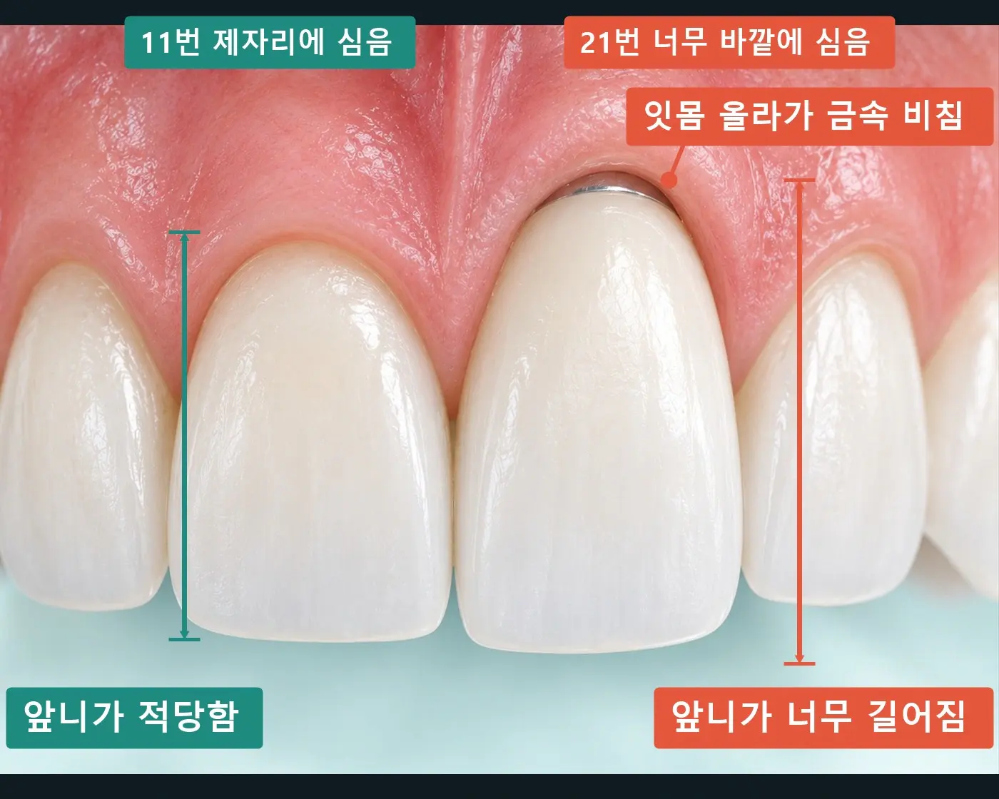
Front-view diagram comparing front tooth #11 at normal length with implant tooth #21 that looks long because the gum has receded

If even one thing is off, only that front tooth looks long

If the axis crosses the incisal edge line, the gum line at that site rises and only the implant tooth looks long. A problem with position or angle shows on the day the prosthesis is attached, and a problem with 2 mm of cheek-side bone shows years later.

Mediolateral cross-section diagram comparing a front tooth implant with the cheek-side gap filled by a bone graft with one without a graft where the cheek-side bone has resorbed and the gum has receded

Place it toward the palate on the day of extraction, and fill the cheek-side gap with a bone graft

If you wait, the thin cheek-side bone resorbs first. So if infection can be removed, the gum has not collapsed, and initial stability is achieved, the implant is placed on the day of extraction. The cheek-side gap created by placing toward the palate is filled with bone graft material to make 2 mm.

Planning Points

The period without front teeth and the number of implants

If initial stability is sufficient, a provisional tooth is connected on the day of placement, and fewer implants than missing teeth are placed.

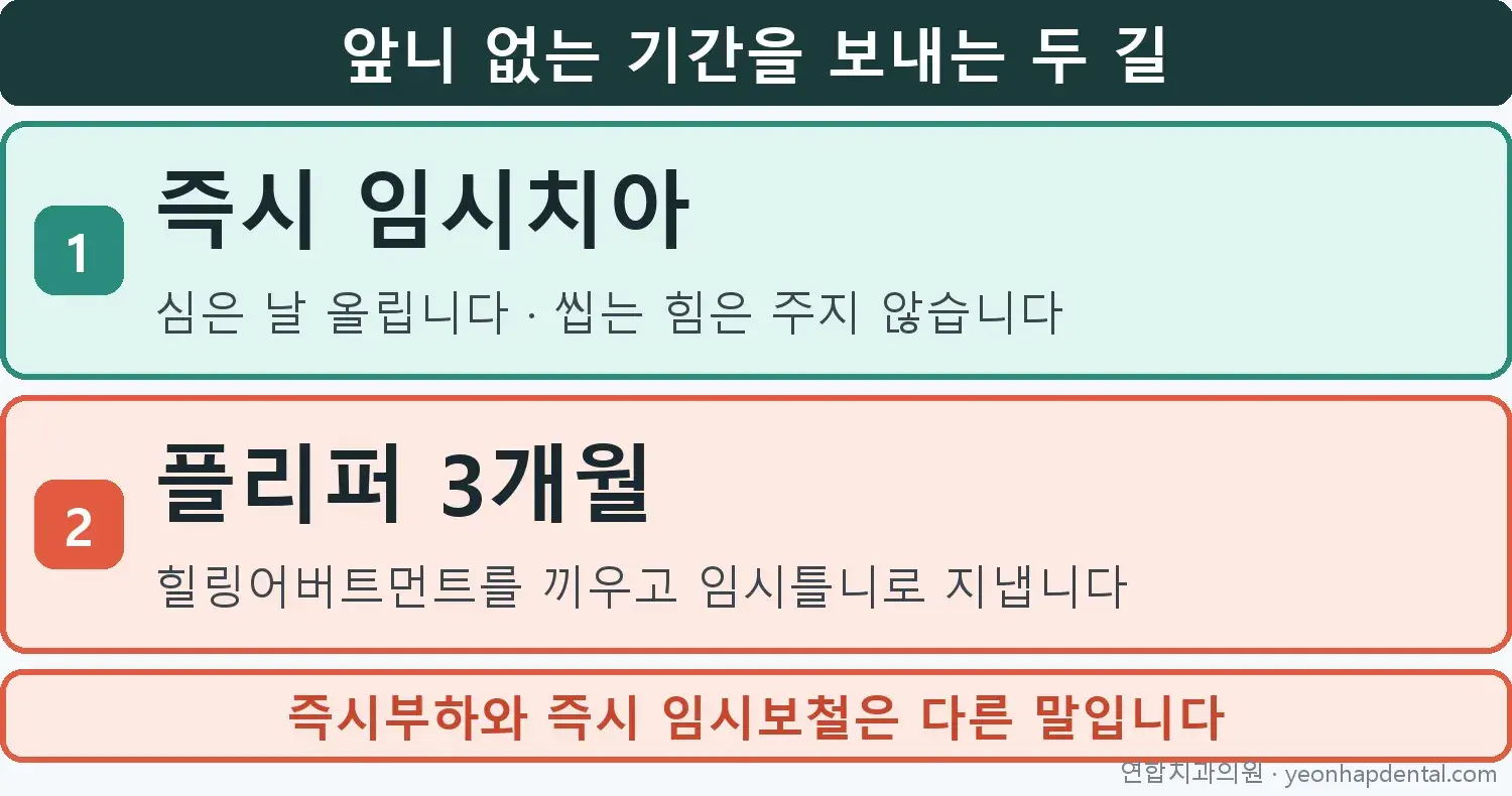
Infographic of two provisional tooth routes for a front tooth implant: an immediate provisional tooth, or a flipper (temporary denture) for 3 months

A provisional tooth on the day of placement, or a flipper (temporary denture) for 3 months

If initial stability is sufficient, a provisional tooth is connected on the day of placement, and it is adjusted so that chewing force does not directly load it. This is different from immediate loading, which is meant for chewing. Depending on the condition of the gum, some patients spend 3 months with a flipper and then move on to the final prosthesis.

Infographic of the number of implants by number of missing front teeth: 3 for the six upper sites, 2 for the six lower sites

Six front tooth sites: 3 implants in the upper jaw, 2 in the lower jaw

If all six upper front tooth sites are missing, we use 3: one at each canine, plus one more at whichever of the four front sites has the better bone; for the six lower front tooth sites, we make it with 2 at the two canines. One site gets 1 implant, and two sites get 1-2 depending on tooth size. If the lateral incisor is small, only one may be placed and joined with a cantilever. The sites are decided on the first-day CT by bone thickness. These are Yeonhap Dental Clinic's planning criteria.

Before Visit

Helpful details before your visit

For front teeth, your smile is the result, so if you let us know the items below, the consultation will be more accurate.

  • Whether you show a lot of gum when you smile. If you have a photo of you smiling, please show us
  • How long ago the tooth was lost, and if it is to be extracted, whether it has ever swollen or discharged pus
  • Whether you take diabetes medication, osteoporosis medication, or medication related to blood clotting
  • Whether you are using a temporary prosthesis such as an existing bridge or flipper

Front tooth implant specifications: Yeonhap Dental Clinic's criteria

Upper front teeth: diameter 3.0-4.0 mm, length 8.5 mm or more. Lower canines: 4.0 mm. Lower middle front teeth: a one-piece implant of 2.5-3.0 mm and 13 mm only when the bone is thin. The standard number is 3 for the six upper front tooth sites and 2 for the six lower front tooth sites. A diameter under 3.0 mm is used only for the lower middle front teeth, where forces are small. This is because it has been reported to have an odds ratio of 4.54 for failure compared with standard diameters (Schiegnitz 2018).
Three things Yeonhap Dental Clinic keeps to for front tooth implants

Problems with front tooth implants show up at two points in time. If the position or angle is wrong, it shows on the day the prosthesis is attached, and if 2 mm of cheek-side bone is lacking, it shows years later as the gum recedes. When the cheek-side bone at placement was under 2 mm, marginal bone loss one year after prosthesis connection is reported as 0.36 mm versus 0.03 mm (prospective study of 39 implants, Nohra 2018), and in immediately placed upper front teeth, the proportion in which the gum receded by more than 1 mm is reported as a median of 26% across 8 studies (range 9-41%, 1 to 3 years) (Chen 2014). For this reason, at Yeonhap Dental Clinic we place the implant toward the palate to secure the cheek-side bone, tilt the axis toward the cheek only within the incisal edge line, and fill the cheek-side gap with a bone graft to make 2 mm. An angle that crosses the line is classified as a danger zone in the literature (Buser 2004), and even if the bone looks ample, Yeonhap Dental Clinic does not place an implant at that angle. References: Chen 2014, systematic review of esthetic outcomes of immediate placement in front teeth · Nohra 2018, marginal bone loss when cheek-side bone is under 2 mm · Buser 2004, danger zone for front tooth implant position · Schiegnitz 2018, meta-analysis of survival of narrow-diameter implants Treatment results may vary depending on each person's oral condition.

Last updated:

Schedule a consultation by phone or Naver Booking

Leave a few details on our consultation page and we'll get back to you promptly.

Frequently Asked Questions

Q. Can a front tooth implant be placed on the day of extraction?
For a front tooth implant, if infection can be removed, the gum has not collapsed, and initial stability is achieved, it is placed on the day of extraction in most cases, because if you wait, the thin cheek-side bone resorbs first. At Yeonhap Dental Clinic, we place the implant toward the palate and fill the gap created on the cheek side with bone graft material to make 2 mm.
Q. Can a front tooth implant make the tooth look long?
If the axis of a front tooth implant goes past the line toward the lip that connects the incisal edges of the neighboring teeth, the gum line can rise and the crown can end up longer than the neighboring teeth. At Yeonhap Dental Clinic, we place it toward the palate, tilt the axis toward the cheek only within that line, and fill the cheek-side gap with a bone graft.
Q. Can a provisional tooth be attached to a front tooth implant on the day of placement?
For a front tooth implant, if initial stability is sufficient, a provisional tooth is connected on the day of placement and adjusted so that chewing force does not directly load it. This is different from immediate loading, which is meant for chewing. Depending on the condition of the gum, some patients spend 3 months with a flipper and then move on to the final prosthesis.
Q. Do front tooth implants use thinner implants than molars?
Front tooth implants use a narrower diameter than molars. For upper front teeth, we use a diameter of 3.0-4.0 mm and a length of 8.5 mm or more; for the middle sites of the lower front teeth, if the bone is thin, we use a one-piece implant of 2.5-3.0 mm in diameter. A diameter under 3.0 mm has been reported to have an odds ratio of 4.54 for failure compared with standard diameters (Schiegnitz 2018), so we use it only in the lower front teeth, where forces are small.