
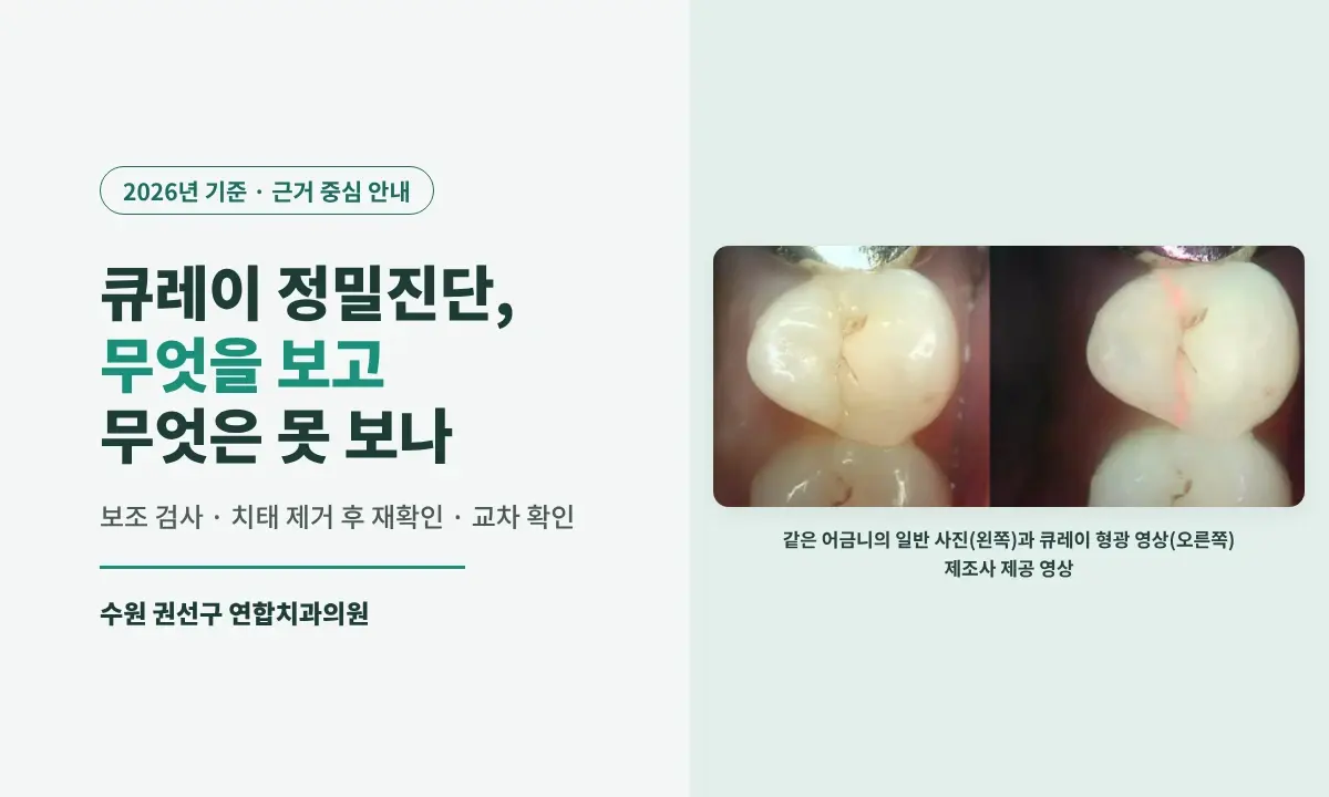
Q-ray shows early cavities, areas around fillings, and suspected cracks using fluorescence. Yeonhap Dental Clinic makes its judgment by adding repeat imaging and checks of radiographs and symptoms.
What does a Q-ray exam check?
You may wonder why we take a Q-ray when we have already looked with our eyes and taken photos. Q-ray shines blue light and captures the fluorescence that comes from the teeth, a way of showing changes that are hard to tell apart by eye.
[Key Answer]
Q-ray is an exam that shows early demineralization (the stage at the start of a cavity when minerals are lost), changes around fillings, and areas where a crack is suspected as fluorescence images. Yeonhap Dental Clinic in Gwonseon-gu, Suwon keeps these images for explanation and for comparison at the next check-up, and makes its judgment by adding repeat imaging, radiographic exams, and a check of your symptoms.
[4-Line Summary]
· Q-ray shows early demineralization, changes around fillings, and suspected cracks by fluorescence, and we keep the images for explanation and for comparison at the next check-up.
· A red reaction can also appear on plaque (the bacterial film on teeth), so we retake images before and after plaque removal and from different angles and compare them.
· Red fluorescence that remains along a crack even after plaque is removed is regarded as an important signal for assessing whether treatment is needed, and we check symptoms, the extent of the crack, and the condition of the nerve together.
· Contacting surfaces between teeth and lesion depth are supplemented with a bitewing radiograph (an X-ray that shows the sides of the molars), and if there is not enough basis for treating now, we compare records over time and watch.
Written by: Dr. Ahn Seong-jin, Chief Director, Yeonhap Dental Clinic
[Table of Contents]
· What does a Q-ray exam check?
· Is everything that looks red on Q-ray a cavity or a crack?
· If red fluorescence remains on a crack, does it need treatment?
· How do we supplement what Q-ray cannot show well?
At Yeonhap Dental Clinic, we take a Q-ray together with regular check-ups. Healthy teeth fluoresce evenly, areas that have lost minerals look dark, and areas with bacterial metabolites look red. This helps find early demineralization that looks fine on the surface, suspected recurrent cavities around fillings, and fine cracks.
The fact that the images are kept is also a major advantage. We can look at the screen together with you and explain where the problem is and why, and at the next check-up we can retake the same area and compare changes.
Studies also report its detection strengths. A meta-analysis pooling the caries detection accuracy of fluorescence exams reported high accuracy in distinguishing sound surfaces from early lesions (Lee 2025). A study of 297 patient teeth showed higher scores than visual and radiographic exams for detecting chewing-surface cavities and cracks (Oh 2021), and in cracked teeth, viewing fluorescence photos together made crack location determination more accurate (Son 2024).
Information checked with Q-ray and how it is used in treatment
| What we check | What shows on Q-ray | How we use it in treatment |
|---|---|---|
| Early demineralization | Areas where fluorescence is dark | Reference for deciding on care and observation before any drilling |
| Changes around fillings | Red or dark reaction along the margin | Check areas where a recurrent cavity is suspected |
| Suspected crack | Dark or red line along the crack | Locate the crack and judge together with symptoms and nerve condition |
| Changes in the same area | Compare side by side with earlier images | Check whether it is progressing over time rather than from a single reading |
Is everything that looks red on Q-ray a cavity or a crack?
Not necessarily. Red fluorescence also comes from plaque and tartar on the tooth surface, so we compare images taken before and after plaque removal and from different angles.
Yeonhap Dental Clinic takes the Q-ray at the start of a regular check-up, before touching the gums. If it cannot be judged at once whether a red reaction is a cavity, a crack, or leftover plaque, we remove the plaque at that site with a probe or similar instrument and then retake the same tooth from a different angle.
Retaking the image gives us information to compare whether the reaction came from surface plaque or remains on the tooth itself. However, we do not confirm a lesion from this comparison alone; we interpret it together with the radiographic exam and symptom check below.
If red fluorescence remains on a crack, does it need treatment?
Red fluorescence that remains along a crack even after plaque is removed is an important signal for assessing whether treatment is needed, and Yeonhap Dental Clinic checks symptoms, the extent of the crack, and the condition of the nerve together to decide between treatment and observation.
We look at three things together. For symptoms, we check whether it hurts when you chew, whether it is sensitive to cold, and whether it hurts even at rest, to gauge the nerve condition. For the extent of the crack, we look at whether it extends past the edge of the side of the tooth. If it hurts even at rest, we test the nerve condition to judge whether root canal treatment is needed, and if it hurts only when chewing, the cause may be something other than the crack, so we sort out the cause first.
This is how our clinic uses it in practice. In a study of cracked teeth, there was no difference in fluorescence values between a cracked tooth with symptoms and the symptom-free cracked tooth on the opposite side (Lee 2023). However, Lee 2023 is not a study that verified whether fluorescence alone can predict the need for treatment. That is why we look at fluorescence together with symptoms, the extent of the crack, and the condition of the nerve. Choosing between resin, inlay, and crown is covered in Cavity Treatment Types: How to Choose Between Resin, Inlay, and Crown.
How do we supplement what Q-ray cannot show well?
Contacting surfaces between teeth and lesion depth are supplemented with a bitewing radiograph, and if there is not enough basis for treating now, we compare records over time and watch.
Where teeth touch, light does not reach evenly, which limits imaging and interpretation. In a study that used bitewings as the reference, the red fluorescence index alone had limited performance in identifying proximal surface cavities (Oh 2021). Adding a bitewing when needed gives information about cavities on contacting surfaces and how deep the lesion is.
If, after all the exams, there is still not enough basis to treat right away, we judge whether watching is appropriate and then, without drilling, retake images under the same conditions at the next regular check-up and compare them with the earlier ones. Because we keep records taken under the same conditions each time, a decision does not have to rest on a single reading.
Limits of Q-ray and how we supplement them (Yeonhap Dental Clinic)
| Limit | Supplementary method | Additional information gained |
|---|---|---|
| Red reaction that can be confused with plaque | Retake before and after plaque removal and from different angles | Whether the reaction is surface plaque or remains on the tooth |
| Limits in assessing contacting surfaces and lesion depth | Bitewing radiograph when needed | Cavities on contacting surfaces and how deep the lesion is |
| Need for treatment of a crack | Check symptoms, extent of the crack, and nerve condition | Basis for deciding whether to treat now or watch |
| Insufficient basis for immediate treatment | Judge whether observation is appropriate, then compare images at the next check-up | Change over time |
4 things to check when consulting about precision diagnosis in Gwonseon-gu and Tap-dong, Suwon
The real standard is not whether the equipment exists but how the results are explained and checked.
Yeonhap Dental Clinic, across from Suwon NongHyup Guun Branch (231 Geumho-ro), on the 5th floor at 230 Geumho-ro, Gwonseon-gu, Suwon, Gyeonggi-do (Tap-dong, Cham Clinic Building), has treated patients in the same place since 2002, and Dr. Ahn Seong-jin, Chief Director and Integrated Dentistry specialist, oversees treatment.
· Whether the exam images are explained on screen together with you
· Whether a red reaction is rechecked after plaque removal
· If it is a crack, whether symptoms, the extent of the crack, and the nerve condition are looked at together
· Whether, when things are unclear, they offer the option of watching and comparing with images from the next check-up
[Key Takeaways]
✓ Q-ray shows early demineralization, changes around fillings, and suspected cracks by fluorescence, and we keep the images for explanation and for comparing progress.
✓ Red fluorescence that remains along a crack after plaque removal is an important signal, and we decide between treatment and observation together with symptoms, the extent of the crack, and nerve condition.
✓ Contacting surfaces and lesion depth are supplemented with a bitewing, and if the basis is insufficient, we compare images at the next check-up.
A Few Words from the Treatment Room About Q-ray Exams
Some patients ask why we take another image when we have already taken photos and looked with our eyes. I tell them it is because each exam shows a different place. When a red reaction appears on Q-ray, I do not reach a conclusion right away; I remove the plaque, look again from a different angle, and check the radiograph and your symptoms together. If it is still unclear, I tell the patient we will look again at the next check-up. You might expect that adding diagnostic equipment leads to more treatment, but my experience has been the opposite. The more exams I overlay, the easier it became to decide to leave alone sites that do not need to be touched. A tooth that has been drilled once does not come back.
References
📋 Treatment Information
This post was written in compliance with the Korean Medical Service Act and the Ministry of Health and Welfare's medical advertising guidelines, and is intended to provide medical information. It does not guarantee or assert the result of any specific exam or treatment. Diagnostic results and treatment direction may vary depending on each person's oral condition, so please be sure to consult fully with your dental team before deciding.
🦷 Precision diagnosis information: Yeonhap Dental Clinic Treatment Departments
📍 5th Floor, 230 Geumho-ro, Gwonseon-gu, Suwon, Gyeonggi-do (Tap-dong, Cham Clinic Building)
🚕 Across from Suwon NongHyup Guun Branch (231 Geumho-ro) / about 15 minutes by taxi from Suwon Station
🚗 Free parking available in the building
📞 031-293-2207
🌐 yeonhapdental.com
🟡 Opened in 2002 / Treatment overseen by Dr. Ahn Seong-jin, Chief Director and Integrated Dentistry specialist
🔗 Online booking and consultation
Q-ray Precision Diagnosis: Frequently Asked Questions
- What does a Q-ray exam check?
- Q-ray is an exam that shines blue light and captures the fluorescence of teeth, showing early demineralization (the stage at the start of a cavity when minerals are lost), changes around fillings, and areas where a crack is suspected. The images are kept, so they can be used for explanation and for comparison at the next check-up.
- If Q-ray shows red, is it a cavity?
- Not necessarily. Red fluorescence also comes from plaque (the bacterial film on teeth) and tartar, so Yeonhap Dental Clinic removes the plaque and then retakes the image from a different angle to compare.
- If Q-ray shows red fluorescence on a crack, do I need a crown?
- Not every visible crack gets a crown. Yeonhap Dental Clinic regards red fluorescence that remains along a crack even after plaque removal as an important signal, and checks symptoms, whether the crack extends past the edge of the side of the tooth, and the nerve condition together to decide between treatment and observation.
- Can Q-ray also show cavities between teeth?
- Where teeth touch, light does not reach evenly, which limits Q-ray imaging and interpretation. When needed, we also check cavities on contacting surfaces and their depth with a bitewing radiograph (an X-ray that shows the sides of the molars).
- If Q-ray shows something abnormal, is it treated right away?
- If there is not enough basis to treat right away, we do not drill and instead watch. Yeonhap Dental Clinic retakes images under the same conditions at the next regular check-up and compares them with the earlier ones.
- Where is Yeonhap Dental Clinic in Tap-dong and Guun-dong, Gwonseon-gu, Suwon?
- We are located on the 5th floor at 230 Geumho-ro, Gwonseon-gu, Suwon, Gyeonggi-do (Tap-dong, Cham Clinic Building), across from Suwon NongHyup Guun Branch (231 Geumho-ro). Free parking is available in the building, and we are about 15 minutes by taxi from Suwon Station.
Related pages
Before your visit
This article provides general information on oral health and dental care. Whether treatment is needed, and which method is appropriate, may change after an oral exam and any necessary diagnostic tests. If you have pain, a loose tooth, swelling, or discomfort with a prosthesis, please visit us for a consultation.