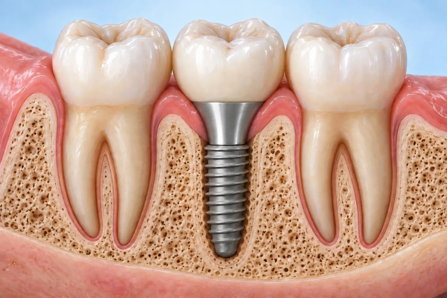
Depth, Placement Direction, and Prosthetic Shape: The Evidence and Its Limits
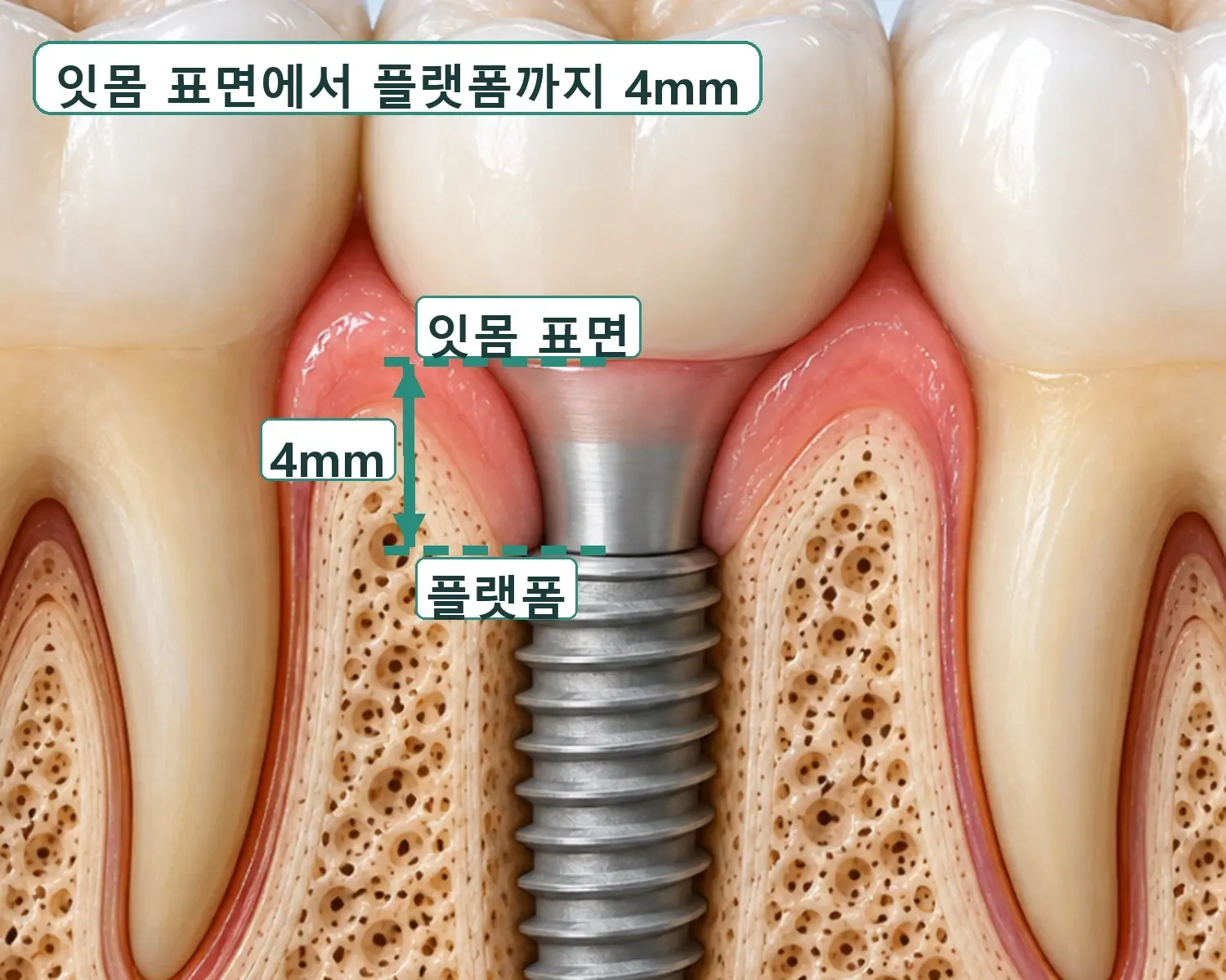
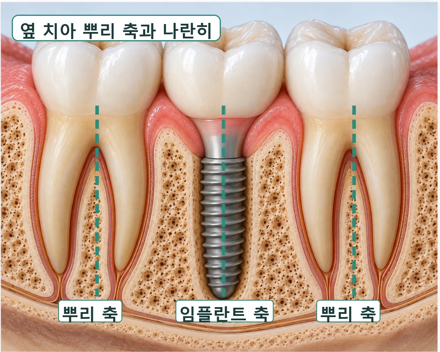
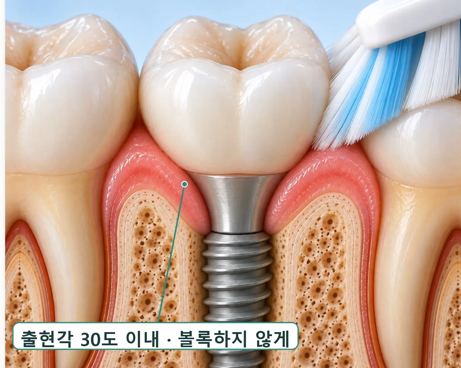
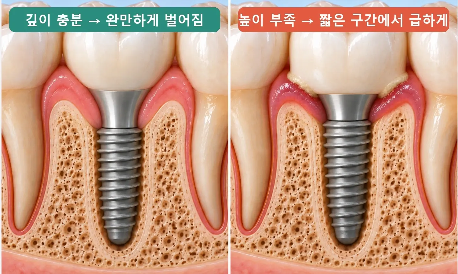
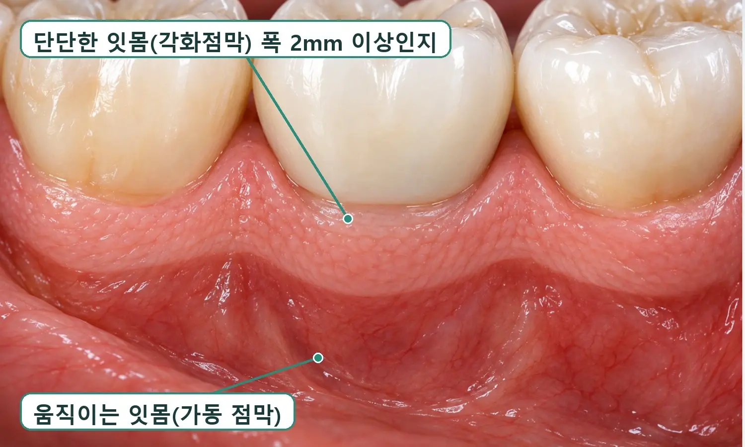
In a study of patients with thin gums, the group placed 1.5 mm below the crest had less marginal bone loss at 2 years than the group placed at crest level with thickened soft tissue (Linkevicius 2020), but early bone changes with deep placement differ from study to study, so this does not mean that deeper is better (Hedayatipanah 2024). In some studies of bone-level implants, peri-implantitis was more frequent with an emergence angle over 30 degrees, particularly when combined with a convex profile, but these were single-time-point comparisons, so causality has not been established (Katafuchi 2018, Soulami 2022). With keratinized mucosa narrower than 2 mm, more plaque, inflammation, and gum recession are reported (Lin 2013). The placement direction criteria are not values from the literature but our own clinic standards.
For this reason, Yeonhap Dental Clinic refers to research findings but decides the actual depth, placement direction, prosthetic shape, and whether to supplement the gum according to each patient's condition.
References: Linkevicius 2020, 2-year randomized study of subcrestal placement in thin gums · Hedayatipanah 2024, vertical implant position and marginal bone loss · Katafuchi 2018, prosthetic emergence angle and profile and peri-implantitis · Soulami 2022, systematic review of emergence angle and profile and peri-implantitis · Lin 2013, systematic review of peri-implant keratinized mucosa
Treatment results may vary by individual oral condition.